National Institutes of Health/National Heart, Lung, and Blood Institute (NIH/NHLBI)
HL140925
United States
National Institutes of Health/National Institute of General Medical Sciences (NIH/NIGMS)
GM116790
United States
National Institutes of Health/National Institute of General Medical Sciences (NIH/NIGMS)
GM116788
United States
Citation
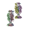
Journal: J Mol Biol / Year: 2021 Title: Interaction of the C2 Ig-like Domain of Cardiac Myosin Binding Protein-C with F-actin. Authors: Cristina M Risi / Malay Patra / Betty Belknap / Samantha P Harris / Howard D White / Vitold E Galkin / Abstract: Cardiac muscle contraction depends on interactions between thick (myosin) and thin (actin) filaments (TFs). TFs are regulated by intracellular Ca levels. Under activating conditions Ca binds to the ...Cardiac muscle contraction depends on interactions between thick (myosin) and thin (actin) filaments (TFs). TFs are regulated by intracellular Ca levels. Under activating conditions Ca binds to the troponin complex and displaces tropomyosin from myosin binding sites on the TF surface to allow actomyosin interactions. Recent studies have shown that in addition to Ca, the first four N-terminal domains (NTDs) of cardiac myosin binding protein C (cMyBP-C) (e.g. C0, C1, M and C2), are potent modulators of the TF activity, but the mechanism of their collective action is poorly understood. Previously, we showed that C1 activates the TF at low Ca and C0 stabilizes binding of C1 to the TF, but the ability of C2 to bind and/or affect the TF remains unknown. Here we obtained 7.5 Å resolution cryo-EM reconstruction of C2-decorated actin filaments to demonstrate that C2 binds to actin in a single structural mode that does not activate the TF unlike the polymorphic binding of C0 and C1 to actin. Comparison of amino acid sequences of C2 with either C0 or C1 shows low levels of identity between the residues involved in interactions with the TF but high levels of conservation for residues involved in Ig fold stabilization. This provides a structural basis for strikingly different interactions of structurally homologous C0, C1 and C2 with the TF. Our detailed analysis of the interaction of C2 with the actin filament provides crucial information required to model the collective action of cMyBP-C NTDs on the cardiac TF.
In the structure databanks used in Yorodumi, some data are registered as the other names, "COVID-19 virus" and "2019-nCoV". Here are the details of the virus and the list of structure data.
Jan 31, 2019. EMDB accession codes are about to change! (news from PDBe EMDB page)
EMDB accession codes are about to change! (news from PDBe EMDB page)
The allocation of 4 digits for EMDB accession codes will soon come to an end. Whilst these codes will remain in use, new EMDB accession codes will include an additional digit and will expand incrementally as the available range of codes is exhausted. The current 4-digit format prefixed with “EMD-” (i.e. EMD-XXXX) will advance to a 5-digit format (i.e. EMD-XXXXX), and so on. It is currently estimated that the 4-digit codes will be depleted around Spring 2019, at which point the 5-digit format will come into force.
The EM Navigator/Yorodumi systems omit the EMD- prefix.
Related info.:Q: What is EMD? / ID/Accession-code notation in Yorodumi/EM Navigator
Yorodumi is a browser for structure data from EMDB, PDB, SASBDB, etc.
This page is also the successor to EM Navigator detail page, and also detail information page/front-end page for Omokage search.
The word "yorodu" (or yorozu) is an old Japanese word meaning "ten thousand". "mi" (miru) is to see.
Related info.:EMDB / PDB / SASBDB / Comparison of 3 databanks / Yorodumi Search / Aug 31, 2016. New EM Navigator & Yorodumi / Yorodumi Papers / Jmol/JSmol / Function and homology information / Changes in new EM Navigator and Yorodumi
 Movie
Movie Controller

 Yorodumi
 Open data
 Basic information
 Components
 Keywords
 Function and homology information
 Homo sapiens (human)
 Authors
 United States, 3items
  Citation
 Structure visualization
 Downloads & links
 Other downloads


 PDBj
PDBj


 Assembly
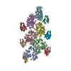

 Sample preparation
 Electron microscopy imaging
 FIELD EMISSION GUN / Accelerating voltage: 300 kV / Illumination mode: FLOOD BEAM
 Processing

