[English] 日本語
Yorodumi
- PDB-5k6p: The NMR structure of the m domain tri-helix bundle and C2 of huma... -

+
Open data


ID or keywords:

Loading...

-
Basic information

Entry
Database: PDB / ID: 5k6p
TitleThe NMR structure of the m domain tri-helix bundle and C2 of human cardiac Myosin Binding Protein C
ComponentsMyosin-binding protein C, cardiac-type
KeywordsCONTRACTILE PROTEIN / immunoglobulin
Function / homology
Function and homology information


C zone / regulation of muscle filament sliding / striated muscle myosin thick filament / cardiac myofibril / regulation of striated muscle contraction / Striated Muscle Contraction / positive regulation of ATP-dependent activity / A band / structural constituent of muscle / ventricular cardiac muscle tissue morphogenesis ...C zone / regulation of muscle filament sliding / striated muscle myosin thick filament / cardiac myofibril / regulation of striated muscle contraction / Striated Muscle Contraction / positive regulation of ATP-dependent activity / A band / structural constituent of muscle / ventricular cardiac muscle tissue morphogenesis / myosin binding / myosin heavy chain binding / ATPase activator activity / heart morphogenesis / titin binding / cardiac muscle contraction / sarcomere / actin binding / cell adhesion / identical protein binding / metal ion binding / cytosol
Similarity search - Function
MyBP-C, tri-helix bundle domain / Tri-helix bundle domain / Immunoglobulin I-set / Immunoglobulin I-set domain / Fibronectin type III domain / Fibronectin type 3 domain / Immunoglobulin subtype 2 / Immunoglobulin C-2 Type / Fibronectin type-III domain profile. / Fibronectin type III ...MyBP-C, tri-helix bundle domain / Tri-helix bundle domain / Immunoglobulin I-set / Immunoglobulin I-set domain / Fibronectin type III domain / Fibronectin type 3 domain / Immunoglobulin subtype 2 / Immunoglobulin C-2 Type / Fibronectin type-III domain profile. / Fibronectin type III / Fibronectin type III superfamily / Immunoglobulin subtype / Immunoglobulin / Ig-like domain profile. / Immunoglobulin-like domain / Immunoglobulin-like domain superfamily / Immunoglobulin-like fold
Similarity search - Domain/homology
Myosin-binding protein C, cardiac-type
Similarity search - Component
Biological speciesHomo sapiens (human)
MethodSOLUTION NMR / molecular dynamics
AuthorsMichie, K.A. / Kwan, A.H. / Tung, C.S. / Guss, J.M. / Trewhella, J.
Funding support Australia, 1items
OrganizationGrant numberCountry
Australian Research Council (ARC)DP120103841 Australia
Citation
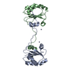
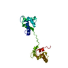
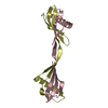
Journal: Structure / Year: 2016
Title: A Highly Conserved Yet Flexible Linker Is Part of a Polymorphic Protein-Binding Domain in Myosin-Binding Protein C.
Authors: Michie, K.A. / Kwan, A.H. / Tung, C.S. / Guss, J.M. / Trewhella, J.
History
DepositionMay 25, 2016Deposition site: RCSB / Processing site: RCSB
Revision 1.0Nov 9, 2016Provider: repository / Type: Initial release
Revision 1.1Nov 16, 2016Group: Database references
Revision 1.2Nov 1, 2017Group: Author supporting evidence / Data collection ...Author supporting evidence / Data collection / Database references / Structure summary
Category: citation / entity ...citation / entity / pdbx_nmr_ensemble / pdbx_struct_assembly_auth_evidence
Item: _citation.journal_id_CSD / _entity.pdbx_number_of_molecules / _pdbx_nmr_ensemble.conformer_selection_criteria
Revision 1.3Jan 1, 2020Group: Author supporting evidence / Data collection / Category: pdbx_audit_support / pdbx_nmr_spectrometer
Item: _pdbx_audit_support.funding_organization / _pdbx_nmr_spectrometer.model
Revision 1.4Jun 14, 2023Group: Database references / Other / Category: database_2 / pdbx_database_status
Item: _database_2.pdbx_DOI / _database_2.pdbx_database_accession / _pdbx_database_status.status_code_nmr_data

-
Structure visualization

Structure viewerMolecule:
MolmilJmol/JSmol

Downloads & links

-
Assembly

Deposited unit
A: Myosin-binding protein C, cardiac-type


Theoretical massNumber of molelcules
Total (without water)15,4981
Polymers15,4981
Non-polymers00
Water0
1


  • Idetical with deposited unit
  • defined by author
  • Evidence: gel filtration, SAXS
TypeNameSymmetry operationNumber
identity operation1_5551
Buried area0 Å2
ΔGint0 kcal/mol
Surface area9170 Å2
NMR ensembles
DataCriteria
Number of conformers (submitted / calculated)20 / 500structures with the lowest energy
RepresentativeModel #1lowest energy
DetailsMonomer as determined by gel filtration and SAXS

-
Components

#1: Protein Myosin-binding protein C, cardiac-type / Cardiac MyBP-C / C-protein / cardiac muscle isoform


Mass: 15497.698 Da / Num. of mol.: 1 / Fragment: residues 319-451
Source method: isolated from a genetically manipulated source
Source: (gene. exp.) Homo sapiens (human) / Tissue: heart / Gene: MYBPC3 / Plasmid: pETM
Details (production host): N-terminal hexahis tag and PreScission protease cleavage site
Production host: Escherichia coli (E. coli) / Strain (production host): Rosetta 2 / References: UniProt: Q14896

-
Experimental details

-
Experiment

ExperimentMethod: SOLUTION NMR
NMR experiment
Conditions-IDExperiment-IDSolution-IDSample stateSpectrometer-IDType
122isotropic23D HN(CA)CB
142isotropic13D HNCO
113isotropic22D 1H-15N HSQC
1163isotropic22D 1H-13C HSQC
133isotropic2HCC(CO)NH TOCSY
183isotropic2CC(CO)NH-TOCSY
173isotropic23D HBHA(CO)NH
193isotropic23D CBCA(CO)NH
1124isotropic23D 1H-15N NOESY
1104isotropic23D 13C NOESY aromatic
164isotropic23D (H)CCH-TOCSY
1204isotropic23D (H)CCH-TOCSY
1155isotropic22D (HB)CB(CGCD)HD
155isotropic12D (HB)CB(CGCDCE)HE
1135isotropic23D 13C NOESY aliphatic
1116isotropic22D 1H-13C HSQC aromatic
1146isotropic23D HCC TOCSY aromatic
1196isotropic23D (H)CCH-TOCSY

-
Sample preparation

Details
TypeSolution-IDContentsLabelSolvent systemDetails
solution2412 uM [U-99% 13C; U-99% 15N] human cMyBP-C (319-451), 10 mM MES, 1 mM CaCl2, 50 mM NaCl, 2 mM TCEP, 1 mM Na azide, 10 mM DSS, 95% H2O/5% D2O15N_13C_sample195% H2O/5% D2O
solution3920 uM [U-99% 13C; U-99% 15N] human cMyBP-C (319-451), 10 mM MES, 1 mM CaCl2, 50 mM NaCl, 2 mM TCEP, 1 mM Na azide, 10 mM DSS, 95% H2O/5% D2O15N_13C_sample295% H2O/5% D2Ohigh concentration sample
solution4550 uM [U-99% 13C; U-99% 15N] human cMyBP-C (319-451), 10 mM MES, 1 mM CaCl2, 50 mM NaCl, 1 mM Na azide, 2 mM TCEP, 10 mM DSS, 95% H2O/5% D2O15N_13C_sample395% H2O/5% D2Odilution of sample 2
solution5500 uM [U-99% 13C; U-99% 15N] human cMyBP-C (319-451), 10 mM MES, 1 mM CaCl2, 50 mM NaCl, 2 mM TCEP, 1 mM Na azide, 10 mM DSS, 99% D2O15N_13C_sample499% D2O15N_13C_sample3, freeze dried (part 1 of 2) and re-suspended in 99% D2O.
solution6800 uM [U-99% 13C; U-99% 15N] human cMyBP-C (319-451), 10 mM MES, 1 mM CaCl2, 50 mM NaCl, 2 mM TCEP, 1 mM Na azide, 10 mM DSS, 99% D2O15N_13C_sample599% D2O15N_13C_sample3 was freeze dried (part 2 of 2) and re-suspended in 99% D2O.
Sample
Conc. (mg/ml)ComponentIsotopic labelingSolution-ID
412 uMhuman cMyBP-C (319-451)[U-99% 13C; U-99% 15N]2
10 mMMESnatural abundance2
1 mMCaCl2natural abundance2
50 mMNaClnatural abundance2
2 mMTCEPnatural abundance2
1 mMNa azidenatural abundance2
10 mMDSSnatural abundance2
920 uMhuman cMyBP-C (319-451)[U-99% 13C; U-99% 15N]3
10 mMMESnatural abundance3
1 mMCaCl2natural abundance3
50 mMNaClnatural abundance3
2 mMTCEPnatural abundance3
1 mMNa azidenatural abundance3
10 mMDSSnatural abundance3
550 uMhuman cMyBP-C (319-451)[U-99% 13C; U-99% 15N]4
10 mMMESnatural abundance4
1 mMCaCl2natural abundance4
50 mMNaClnatural abundance4
1 mMNa azidenatural abundance4
2 mMTCEPnatural abundance4
10 mMDSSnatural abundance4
500 uMhuman cMyBP-C (319-451)[U-99% 13C; U-99% 15N]5
10 mMMESnatural abundance5
1 mMCaCl2natural abundance5
50 mMNaClnatural abundance5
2 mMTCEPnatural abundance5
1 mMNa azidenatural abundance5
10 mMDSSnatural abundance5
800 uMhuman cMyBP-C (319-451)[U-99% 13C; U-99% 15N]6
10 mMMESnatural abundance6
1 mMCaCl2natural abundance6
50 mMNaClnatural abundance6
2 mMTCEPnatural abundance6
1 mMNa azidenatural abundance6
10 mMDSSnatural abundance6
Sample conditionsIonic strength: 50 mM / Label: std_condition / pH: 6.5 / Pressure: 1 atm / Temperature: 298 K

-
NMR measurement

NMR spectrometer
TypeManufacturerModelField strength (MHz)Spectrometer-ID
Bruker AVANCEBrukerAVANCE6001
Bruker AVANCEBrukerAVANCE8002

-
Processing

NMR software
NameVersionDeveloperClassification
CYANAGuntert, Mumenthaler and Wuthrichrefinement
CNS1.2Brunger, Adams, Clore, Gros, Nilges and Readrefinement
AnalysisCCPNchemical shift assignment
AnalysisCCPNpeak picking
CYANAGuntert, Mumenthaler and Wuthrichstructure calculation
RefinementMethod: molecular dynamics / Software ordinal: 2
Details: torsion angle dynamics, water refinement was carried out using the lowest 50 energy structures from CYANA
NMR representativeSelection criteria: lowest energy
NMR ensembleConformer selection criteria: structures with the lowest energy
Conformers calculated total number: 500 / Conformers submitted total number: 20

+
About Yorodumi

-
News

-
Feb 9, 2022. New format data for meta-information of EMDB entries

New format data for meta-information of EMDB entries

  • Version 3 of the EMDB header file is now the official format.
  • The previous official version 1.9 will be removed from the archive.

Related info.:EMDB header

External links:wwPDB to switch to version 3 of the EMDB data model

-
Aug 12, 2020. Covid-19 info

Covid-19 info

URL: https://pdbj.org/emnavi/covid19.php

New page: Covid-19 featured information page in EM Navigator.

Related info.:Covid-19 info / Mar 5, 2020. Novel coronavirus structure data

+
Mar 5, 2020. Novel coronavirus structure data

Novel coronavirus structure data

Related info.:Yorodumi Speices / Aug 12, 2020. Covid-19 info

External links:COVID-19 featured content - PDBj / Molecule of the Month (242):Coronavirus Proteases

+
Jan 31, 2019. EMDB accession codes are about to change! (news from PDBe EMDB page)

EMDB accession codes are about to change! (news from PDBe EMDB page)

  • The allocation of 4 digits for EMDB accession codes will soon come to an end. Whilst these codes will remain in use, new EMDB accession codes will include an additional digit and will expand incrementally as the available range of codes is exhausted. The current 4-digit format prefixed with “EMD-” (i.e. EMD-XXXX) will advance to a 5-digit format (i.e. EMD-XXXXX), and so on. It is currently estimated that the 4-digit codes will be depleted around Spring 2019, at which point the 5-digit format will come into force.
  • The EM Navigator/Yorodumi systems omit the EMD- prefix.

Related info.:Q: What is EMD? / ID/Accession-code notation in Yorodumi/EM Navigator

External links:EMDB Accession Codes are Changing Soon! / Contact to PDBj

+
Jul 12, 2017. Major update of PDB

Major update of PDB

  • wwPDB released updated PDB data conforming to the new PDBx/mmCIF dictionary.
  • This is a major update changing the version number from 4 to 5, and with Remediation, in which all the entries are updated.
  • In this update, many items about electron microscopy experimental information are reorganized (e.g. em_software).
  • Now, EM Navigator and Yorodumi are based on the updated data.

External links:wwPDB Remediation / Enriched Model Files Conforming to OneDep Data Standards Now Available in the PDB FTP Archive

-
Yorodumi

Thousand views of thousand structures

  • Yorodumi is a browser for structure data from EMDB, PDB, SASBDB, etc.
  • This page is also the successor to EM Navigator detail page, and also detail information page/front-end page for Omokage search.
  • The word "yorodu" (or yorozu) is an old Japanese word meaning "ten thousand". "mi" (miru) is to see.

Related info.:EMDB / PDB / SASBDB / Comparison of 3 databanks / Yorodumi Search / Aug 31, 2016. New EM Navigator & Yorodumi / Yorodumi Papers / Jmol/JSmol / Function and homology information / Changes in new EM Navigator and Yorodumi

Read more