National Institutes of Health/National Institute of General Medical Sciences (NIH/NIGMS)
GM118326
United States
Citation
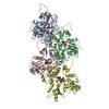
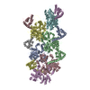
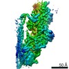
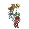
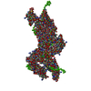
Journal: Elife / Year: 2020 Title: Structural basis of αE-catenin-F-actin catch bond behavior. Authors: Xiao-Ping Xu / Sabine Pokutta / Megan Torres / Mark F Swift / Dorit Hanein / Niels Volkmann / William I Weis / Abstract: Cell-cell and cell-matrix junctions transmit mechanical forces during tissue morphogenesis and homeostasis. α-Catenin links cell-cell adhesion complexes to the actin cytoskeleton, and mechanical ...Cell-cell and cell-matrix junctions transmit mechanical forces during tissue morphogenesis and homeostasis. α-Catenin links cell-cell adhesion complexes to the actin cytoskeleton, and mechanical load strengthens its binding to F-actin in a direction-sensitive manner. Specifically, optical trap experiments revealed that force promotes a transition between weak and strong actin-bound states. Here, we describe the cryo-electron microscopy structure of the F-actin-bound αE-catenin actin-binding domain, which in solution forms a five-helix bundle. In the actin-bound structure, the first helix of the bundle dissociates and the remaining four helices and connecting loops rearrange to form the interface with actin. Deletion of the first helix produces strong actin binding in the absence of force, suggesting that the actin-bound structure corresponds to the strong state. Our analysis explains how mechanical force applied to αE-catenin or its homolog vinculin favors the strongly bound state, and the dependence of catch bond strength on the direction of applied force.
History
Deposition
May 6, 2020
Deposition site: RCSB / Processing site: RCSB
Revision 1.0
Oct 7, 2020
Provider: repository / Type: Initial release
Revision 1.0
Oct 7, 2020
Data content type: EM metadata / Data content type: EM metadata / Provider: repository / Type: Initial release
Revision 1.0
Oct 7, 2020
Data content type: Image / Data content type: Image / Provider: repository / Type: Initial release
Revision 1.0
Oct 7, 2020
Data content type: Primary map / Data content type: Primary map / Provider: repository / Type: Initial release
Data content type: EM metadata Data content type: EM metadata / EM metadata ...EM metadata / EM metadata / EM metadata / EM metadata Group: Data processing / Database references ...Data processing / Database references / Experimental summary / Refinement description Data content type: EM metadata / EM metadata ...EM metadata / EM metadata / EM metadata / EM metadata Category: database_2 / em_3d_fitting_list ...database_2 / em_3d_fitting_list / em_admin / pdbx_initial_refinement_model Data content type: EM metadata / EM metadata ...EM metadata / EM metadata / EM metadata / EM metadata / EM metadata / EM metadata / EM metadata Item: _database_2.pdbx_DOI / _database_2.pdbx_database_accession ..._database_2.pdbx_DOI / _database_2.pdbx_database_accession / _em_3d_fitting_list.accession_code / _em_3d_fitting_list.initial_refinement_model_id / _em_3d_fitting_list.source_name / _em_3d_fitting_list.type / _em_admin.last_update
Average exposure time: 1 sec. / Electron dose: 60 e/Å2 / Detector mode: INTEGRATING / Film or detector model: FEI FALCON II (4k x 4k) / Num. of grids imaged: 3 / Num. of real images: 5573
Image scans
Sampling size: 14 µm / Width: 4096 / Height: 4096 / Movie frames/image: 7 / Used frames/image: 2-7
In the structure databanks used in Yorodumi, some data are registered as the other names, "COVID-19 virus" and "2019-nCoV". Here are the details of the virus and the list of structure data.
Jan 31, 2019. EMDB accession codes are about to change! (news from PDBe EMDB page)
EMDB accession codes are about to change! (news from PDBe EMDB page)
The allocation of 4 digits for EMDB accession codes will soon come to an end. Whilst these codes will remain in use, new EMDB accession codes will include an additional digit and will expand incrementally as the available range of codes is exhausted. The current 4-digit format prefixed with “EMD-” (i.e. EMD-XXXX) will advance to a 5-digit format (i.e. EMD-XXXXX), and so on. It is currently estimated that the 4-digit codes will be depleted around Spring 2019, at which point the 5-digit format will come into force.
The EM Navigator/Yorodumi systems omit the EMD- prefix.
Related info.:Q: What is EMD? / ID/Accession-code notation in Yorodumi/EM Navigator
Yorodumi is a browser for structure data from EMDB, PDB, SASBDB, etc.
This page is also the successor to EM Navigator detail page, and also detail information page/front-end page for Omokage search.
The word "yorodu" (or yorozu) is an old Japanese word meaning "ten thousand". "mi" (miru) is to see.
Related info.:EMDB / PDB / SASBDB / Comparison of 3 databanks / Yorodumi Search / Aug 31, 2016. New EM Navigator & Yorodumi / Yorodumi Papers / Jmol/JSmol / Function and homology information / Changes in new EM Navigator and Yorodumi
 Movie
Movie Controller


 Open data
 Basic information
 Components
 Keywords
 Function and homology information
 Authors
 United States, 1items
  Citation
 Structure visualization
 Downloads & links
 Other downloads
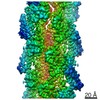


 PDBj
PDBj


 Assembly


 Mass: 24.305 Da / Num. of mol.: 6 / Source method: obtained synthetically / Formula: Mg
 Mass: 427.201 Da / Num. of mol.: 6 / Source method: obtained synthetically / Formula: C10H15N5O10P2 / Comment: ADP, energy-carrying molecule*YM
 Sample preparation
 Electron microscopy imaging
 FIELD EMISSION GUN / Accelerating voltage: 300 kV / Illumination mode: FLOOD BEAM
 Processing