- PDB-6i7s: Microsomal triglyceride transfer protein -
+
Open data
ID or keywords:
Loading...
-
Basic information
Entry
Database: PDB / ID: 6i7s
Title
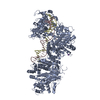
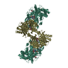
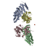
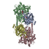
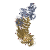
Microsomal triglyceride transfer protein
Components
Microsomal triglyceride transfer protein large subunit
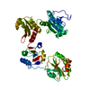
Protein disulfide-isomerase
Keywords
LIPID TRANSPORT / Lipid transfer / protein complex / protein disulfide isomerase
Function / homology
Function and homology information
plasma lipoprotein particle assembly / triglyceride transfer activity / chylomicron assembly / peptidyl-proline hydroxylation to 4-hydroxy-L-proline / regulation of oxidative stress-induced intrinsic apoptotic signaling pathway / phosphatidylcholine transfer activity / procollagen-proline 4-dioxygenase complex / triglyceride transport / insulin processing / phosphatidylethanolamine transfer activity ...plasma lipoprotein particle assembly / triglyceride transfer activity / chylomicron assembly / peptidyl-proline hydroxylation to 4-hydroxy-L-proline / regulation of oxidative stress-induced intrinsic apoptotic signaling pathway / phosphatidylcholine transfer activity / procollagen-proline 4-dioxygenase complex / triglyceride transport / insulin processing / phosphatidylethanolamine transfer activity / VLDL assembly / thiol oxidase activity / procollagen-proline 4-dioxygenase activity / phospholipid transfer activity / LDL remodeling / ceramide 1-phosphate transfer activity / protein disulfide-isomerase / very-low-density lipoprotein particle assembly / endoplasmic reticulum chaperone complex / : / protein folding in endoplasmic reticulum / Collagen biosynthesis and modifying enzymes / lipid carrier activity / Chylomicron assembly / lipoprotein metabolic process / phospholipid transport / Interleukin-23 signaling / interleukin-23-mediated signaling pathway / cholesterol transfer activity / interleukin-12-mediated signaling pathway / Interleukin-12 signaling / cellular response to interleukin-7 / Insulin processing / Detoxification of Reactive Oxygen Species / protein disulfide isomerase activity / microvillus membrane / endoplasmic reticulum-Golgi intermediate compartment / protein-disulfide reductase activity / apolipoprotein binding / protein secretion / endoplasmic reticulum to Golgi vesicle-mediated transport / positive regulation of T cell migration / positive regulation of substrate adhesion-dependent cell spreading / positive regulation of cell adhesion / response to endoplasmic reticulum stress / cholesterol homeostasis / Post-translational protein phosphorylation / lipid metabolic process / brush border membrane / circadian rhythm / Hedgehog ligand biogenesis / response to calcium ion / integrin binding / Regulation of Insulin-like Growth Factor (IGF) transport and uptake by Insulin-like Growth Factor Binding Proteins (IGFBPs) / melanosome / lamellipodium / protein folding / actin binding / cellular response to hypoxia / vesicle / basolateral plasma membrane / positive regulation of viral entry into host cell / cytoskeleton / signaling receptor complex / endoplasmic reticulum lumen / protein heterodimerization activity / external side of plasma membrane / focal adhesion / lipid binding / protein-containing complex binding / enzyme binding / Golgi apparatus / endoplasmic reticulum / protein-containing complex / RNA binding / extracellular exosome / extracellular region / cytosol Similarity search - Function
Lipovitellin-phosvitin complex; beta-sheet shell regions / Lipovitellin; beta-sheet shell regions, chain A / Microsomal triglyceride transfer protein large subunit / MTP large subunit, lipid-binding domain / MTP large subunit, lipid-binding domain / Vitellogenin, N-terminal / Lipovitellin-phosvitin complex, superhelical domain / Vitellinogen, beta-sheet N-terminal / Lipid transport protein, beta-sheet shell / Vitellogenin TPR domain ...Lipovitellin-phosvitin complex; beta-sheet shell regions / Lipovitellin; beta-sheet shell regions, chain A / Microsomal triglyceride transfer protein large subunit / MTP large subunit, lipid-binding domain / MTP large subunit, lipid-binding domain / Vitellogenin, N-terminal / Lipovitellin-phosvitin complex, superhelical domain / Vitellinogen, beta-sheet N-terminal / Lipid transport protein, beta-sheet shell / Vitellogenin TPR domain / Vitellogenin domain profile. / Lipoprotein N-terminal Domain / Protein disulphide isomerase / Thioredoxin-like domain / Disulphide isomerase / Endoplasmic reticulum targeting sequence. / Thioredoxin / Thioredoxin family active site. / Thioredoxin, conserved site / Thioredoxin domain profile. / Thioredoxin domain / Glutaredoxin / Glutaredoxin / Thioredoxin-like superfamily / Roll / 3-Layer(aba) Sandwich / Mainly Beta / Alpha Beta Similarity search - Domain/homology
DI(HYDROXYETHYL)ETHER / TRIETHYLENE GLYCOL / Protein disulfide-isomerase / Microsomal triglyceride transfer protein large subunit Similarity search - Component
A: Protein disulfide-isomerase B: Protein disulfide-isomerase G: Microsomal triglyceride transfer protein large subunit H: Microsomal triglyceride transfer protein large subunit hetero molecules
In the structure databanks used in Yorodumi, some data are registered as the other names, "COVID-19 virus" and "2019-nCoV". Here are the details of the virus and the list of structure data.
Jan 31, 2019. EMDB accession codes are about to change! (news from PDBe EMDB page)
EMDB accession codes are about to change! (news from PDBe EMDB page)
The allocation of 4 digits for EMDB accession codes will soon come to an end. Whilst these codes will remain in use, new EMDB accession codes will include an additional digit and will expand incrementally as the available range of codes is exhausted. The current 4-digit format prefixed with “EMD-” (i.e. EMD-XXXX) will advance to a 5-digit format (i.e. EMD-XXXXX), and so on. It is currently estimated that the 4-digit codes will be depleted around Spring 2019, at which point the 5-digit format will come into force.
The EM Navigator/Yorodumi systems omit the EMD- prefix.
Related info.:Q: What is EMD? / ID/Accession-code notation in Yorodumi/EM Navigator
Yorodumi is a browser for structure data from EMDB, PDB, SASBDB, etc.
This page is also the successor to EM Navigator detail page, and also detail information page/front-end page for Omokage search.
The word "yorodu" (or yorozu) is an old Japanese word meaning "ten thousand". "mi" (miru) is to see.
Related info.:EMDB / PDB / SASBDB / Comparison of 3 databanks / Yorodumi Search / Aug 31, 2016. New EM Navigator & Yorodumi / Yorodumi Papers / Jmol/JSmol / Function and homology information / Changes in new EM Navigator and Yorodumi
 Movie
Movie Controller


 Open data
 Basic information
 Components
 Keywords
 Function and homology information
 Homo sapiens (human)
 X-RAY DIFFRACTION /
  Authors
 Finland, 2items
  Citation
 Structure visualization
 Downloads & links
 Other downloads
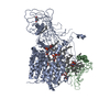


 PDBj
PDBj


 Assembly
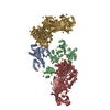


 Mass: 40.078 Da / Num. of mol.: 2 / Source method: obtained synthetically / Formula: Ca
 Mass: 62.068 Da / Num. of mol.: 19 / Source method: obtained synthetically / Formula: C2H6O2
 Mass: 150.173 Da / Num. of mol.: 6 / Source method: obtained synthetically / Formula: C6H14O4
 Mass: 96.063 Da / Num. of mol.: 11 / Source method: obtained synthetically / Formula: SO4
 Mass: 106.120 Da / Num. of mol.: 9 / Source method: obtained synthetically / Formula: C4H10O3
 Mass: 398.489 Da / Num. of mol.: 2 / Source method: obtained synthetically / Formula: C18H38O9 / Comment: precipitant*YM
 Mass: 122.143 Da / Num. of mol.: 1 / Source method: obtained synthetically / Formula: C4H12NO3 / Comment: pH buffer*YM
 Mass: 354.436 Da / Num. of mol.: 1 / Source method: obtained synthetically / Formula: C16H34O8 / Comment: precipitant*YM
 Sample preparation
 / Beamline: MASSIF-1 / Wavelength: 0.966 Å
 Processing