 Movie
Movie Controller


+ Open data

- Basic information
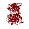
| Entry | Database: PDB / ID: 1lsh | |||||||||
|---|---|---|---|---|---|---|---|---|---|---|
| Title | LIPID-PROTEIN INTERACTIONS IN LIPOVITELLIN | |||||||||
 Components Components |
| |||||||||
 Keywords Keywords | LIPID BINDING PROTEIN / LIPOVITELLIN / VITELLOGENIN / LIPOPROTEIN / PLASMA APOLIPOPROTE APOLIPOPROTEIN B / APOB / MICROSOMAL TRIGLYCERIDE TRANSFER PR BOUNDARY LIPID / PHOSPHOLIPID STRUCTURE | |||||||||
| Function / homology |  Function and homology information Function and homology informationnutrient reservoir activity / lipid carrier activity / cellular response to estrogen stimulus / response to estradiol Similarity search - Function | |||||||||
| Biological species |  Ichthyomyzon unicuspis (silver lamprey) Ichthyomyzon unicuspis (silver lamprey) | |||||||||
| Method |  X-RAY DIFFRACTION / SYNCHROTRON / FOURIER SYNTHESIS / Resolution: 1.9 Å X-RAY DIFFRACTION / SYNCHROTRON / FOURIER SYNTHESIS / Resolution: 1.9 Å | |||||||||
 Authors Authors | Thompson, J.R. / Banaszak, L.J. | |||||||||
 Citation Citation | Journal: Biochemistry / Year: 2002 Title: Lipid-protein interactions in lipovitellin. Authors: Thompson, J.R. / Banaszak, L.J. #1: Journal: Structure / Year: 1998Title: The Structural Basis of Lipid Interactions in Lipovitellin, a Souble Lipoprotein Authors: Anderson, T.A. / Levitt, D.G. / Banaszak, L.J. | |||||||||
| History |
| |||||||||
| Remark 99 | The disulfide bond S-S involving residues 156 and 182 in chain A is formed between the alternate ...The disulfide bond S-S involving residues 156 and 182 in chain A is formed between the alternate conformers B of these residues. | |||||||||
| Remark 600 | HETEROGEN DUE TO LACK OF ELECTRON DENSITY, ONLY VARYING PARTS OF PHOSPHOLIPID MOLECULES COULD BE ...HETEROGEN DUE TO LACK OF ELECTRON DENSITY, ONLY VARYING PARTS OF PHOSPHOLIPID MOLECULES COULD BE MODELED. THE FIRST SEVEN MOLECULES WERE MODELED AS PHOSPHATIDYLCHOLINE WITH AN ALTERNATE CONFORMATION FOR ONE OF THE ACYL CHAINS IN MOLECULES 1 AND 5. MOLECULES 2-7 WERE MODELED AS UNKNOWN PHOSPHOLIPID BUT ARBITARILY CALLED PHOSPHATIDYLCHOLINE. ADDITIONAL 43 METHYLENE CHAIN FRAGMENTS WITH BETWEEN 20 AND 5 CARBON ATOMS EACH WERE MODELED. THREE OF THE HYDROCARBON SEGMENTS ARE BRANCHED BUT NOT MODELED AS PHOSPHOLIPID BECAUSE IT WAS IMPOSSIBLE TO IDENTIFY WHICH PORTION OF ELECTRON DENSITY BELONGED TO THE ACYL CHAINS AND POLAR HEADGROUPS. THE 43 MOLECULES NAMED UPL (ARBITARILY CALLED UNKNOWN BRANCHED PHOSPHOLIPID FRAGMENT) WAS SIMPLY MEANT TO REPRESENTS METHYLENE CHAIN FRAGMENTS. | |||||||||
| Remark 999 | SEQUENCE LIPOVITELLIN IS PROTEOLYTICALLY PROCESSED FROM ITS PRECURSOR PRODUCING FOUR POLYPEPTIDES. ...SEQUENCE LIPOVITELLIN IS PROTEOLYTICALLY PROCESSED FROM ITS PRECURSOR PRODUCING FOUR POLYPEPTIDES. PRECISE CLEAVED LENGTHS OF FRAGMENTS LV-1N, LV-1C AND LV-2 ARE NOT KNOWN. BOTH LV-1N AND LV-1C ARE PRESENTED HERE AS SINGLE ENTITY WITH CHAIN A TO CORRESSPOND WITH HOMOGENEOUS SINGLE POLYPEPTIDE IN LIPOVITELLINS OF OTHER SPECIES. ONE OF THE FRAGMENTS, PHOVITIN CONSISTING OF 232 RESIDUES (1074-1305) IN THE SWS ENTRY WAS PRESENT IN THE CRYSTAL BUT NONE OF ITS RESIDUE COULD BE MODELED DUE TO LACK OF ELECTRON DENSITY. |
- Structure visualization
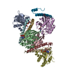
| Structure viewer | Molecule: MolmilJmol/JSmol |
|---|
- Downloads & links
-Download
| PDBx/mmCIF format | 1lsh.cif.gz | 309.5 KB | Display | PDBx/mmCIF format |
|---|---|---|---|---|
| PDB format | pdb1lsh.ent.gz | 230.6 KB | Display | PDB format |
| PDBx/mmJSON format | 1lsh.json.gz | Tree view | PDBx/mmJSON format | |
| Others |  Other downloads Other downloads |
-Validation report
| Arichive directory | https://data.pdbj.org/pub/pdb/validation_reports/ls/1lshftp://data.pdbj.org/pub/pdb/validation_reports/ls/1lsh | HTTPS FTP |
|---|
-Related structure data
| Similar structure data |
|---|


-Links
 PDBj
PDBj- Assembly
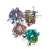
| Deposited unit | 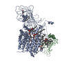
| ||||||||
|---|---|---|---|---|---|---|---|---|---|
| 1 |
| ||||||||
| Unit cell |
| ||||||||
| Details | The biological assembly is a dimer believed to be generated by the crystallographic c2 dyad -x,y,-z |
-Components
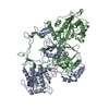
| #1: Protein | Mass: 117469.508 Da / Num. of mol.: 1 / Fragment: LV1n, LV1c / Source method: isolated from a natural source / Details: Egg Yolk / Source: (natural) Ichthyomyzon unicuspis (silver lamprey) / References: UniProt: Q91062 | ||||||
|---|---|---|---|---|---|---|---|
| #2: Protein | Mass: 35110.094 Da / Num. of mol.: 1 / Fragment: LV2 / Source method: isolated from a natural source / Details: Egg Yolk / Source: (natural) Ichthyomyzon unicuspis (silver lamprey) / References: UniProt: Q91062 | ||||||
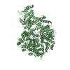
| #3: Chemical | ChemComp-PLD /   Mass: 875.313 Da / Num. of mol.: 7 / Source method: obtained synthetically / Formula: C50H101NO8P Mass: 875.313 Da / Num. of mol.: 7 / Source method: obtained synthetically / Formula: C50H101NO8P#4: Chemical | ChemComp-UPL / 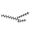  Mass: 478.920 Da / Num. of mol.: 43 / Source method: obtained synthetically / Formula: C34H70 Mass: 478.920 Da / Num. of mol.: 43 / Source method: obtained synthetically / Formula: C34H70#5: Water | ChemComp-HOH / |  Mass: 18.015 Da / Num. of mol.: 1122 / Source method: isolated from a natural source / Formula: H2O Mass: 18.015 Da / Num. of mol.: 1122 / Source method: isolated from a natural source / Formula: H2OHas protein modification | Y | |
-Experimental details
-Experiment
| Experiment | Method: X-RAY DIFFRACTION / Number of used crystals: 1 |
|---|
- Sample preparation
| Crystal | Density Matthews: 2.32 Å3/Da / Density % sol: 46.94 % | ||||||||||||||||||||||||||||||||||||||||||
|---|---|---|---|---|---|---|---|---|---|---|---|---|---|---|---|---|---|---|---|---|---|---|---|---|---|---|---|---|---|---|---|---|---|---|---|---|---|---|---|---|---|---|---|
| Crystal grow | Temperature: 291 K / Method: vapor diffusion, hanging drop / pH: 6.2 Details: Protein stock:10 mg/ml lipovitellin, 0.25 mM sodium citrate; Well: 0.55-0.61 M sodium citrate, 1mM thioglycerol, 1 mM EDTA, 0.05% sodium azide, pH 6.2, VAPOR DIFFUSION, HANGING DROP, temperature 291K | ||||||||||||||||||||||||||||||||||||||||||
| Crystal grow | *PLUS Temperature: 18 ℃ | ||||||||||||||||||||||||||||||||||||||||||
| Components of the solutions | *PLUS
|
-Data collection
| Diffraction | Mean temperature: 100 K |
|---|---|
| Diffraction source | Source: SYNCHROTRON / Site: APS  / Beamline: 19-ID / Wavelength: 1.0332 Å / Beamline: 19-ID / Wavelength: 1.0332 Å |
| Detector | Type: CUSTOM-MADE / Detector: CCD / Date: Feb 7, 1998 / Details: vertically focusing mirror |
| Radiation | Monochromator: double crystal Si220 monochromator,double crystal Si111 monochromator Protocol: SINGLE WAVELENGTH / Monochromatic (M) / Laue (L): M / Scattering type: x-ray |
| Radiation wavelength | Wavelength: 1.0332 Å / Relative weight: 1 |
| Reflection | Resolution: 1.9→30 Å / Num. all: 82007 / Num. obs: 82007 / % possible obs: 74.4 % / Observed criterion σ(F): 0 / Observed criterion σ(I): 0 / Redundancy: 1.54 % / Biso Wilson estimate: 23.3 Å2 / Rsym value: 0.064 / Net I/σ(I): 12.2 |
| Reflection shell | Resolution: 1.9→1.99 Å / % possible all: 30.1 |
| Reflection | *PLUS Lowest resolution: 30 Å / Rmerge(I) obs: 0.064 |
- Processing
| Software |
| ||||||||||||||||||||||||||||||||||||
|---|---|---|---|---|---|---|---|---|---|---|---|---|---|---|---|---|---|---|---|---|---|---|---|---|---|---|---|---|---|---|---|---|---|---|---|---|---|
| Refinement | Method to determine structure: FOURIER SYNTHESIS Starting model: LIPOVITELLIN (ROOM TEMP) Resolution: 1.9→21.6 Å / Rfactor Rfree error: 0.008 / Isotropic thermal model: RESTRAINED / Cross valid method: THROUGHOUT / σ(F): 0 / Stereochemistry target values: Engh & Huber Details: Used OUTIER program (Randy Read) to reject 312 reflections in the range 30-5.9 Angstrom
| ||||||||||||||||||||||||||||||||||||
| Solvent computation | Solvent model: FLAT MODEL / Bsol: 180.794 Å2 / ksol: 0.383954 e/Å3 | ||||||||||||||||||||||||||||||||||||
| Displacement parameters | Biso mean: 49 Å2
| ||||||||||||||||||||||||||||||||||||
| Refine analyze |
| ||||||||||||||||||||||||||||||||||||
| Refinement step | Cycle: LAST / Resolution: 1.9→21.6 Å
| ||||||||||||||||||||||||||||||||||||
| Refine LS restraints |
| ||||||||||||||||||||||||||||||||||||
| LS refinement shell | Resolution: 1.9→1.99 Å / Rfactor Rfree error: 0.072 / Total num. of bins used: 8
| ||||||||||||||||||||||||||||||||||||
| Xplor file |
| ||||||||||||||||||||||||||||||||||||
| Software | *PLUS Version: 0.9,1.0,1.1 / Classification: refinement | ||||||||||||||||||||||||||||||||||||
| Refinement | *PLUS Highest resolution: 1.9 Å / Lowest resolution: 30 Å | ||||||||||||||||||||||||||||||||||||
| Solvent computation | *PLUS | ||||||||||||||||||||||||||||||||||||
| Displacement parameters | *PLUS | ||||||||||||||||||||||||||||||||||||
| Refine LS restraints | *PLUS
|
