 Movie
Movie Controller

[English] 日本語
 Yorodumi
- EMDB-7462: Insulin Receptor ectodomain in complex with two insulin molecules -
+ Open data

- Basic information
| Entry | Database: EMDB / ID: EMD-7462 | ||||||||||||
|---|---|---|---|---|---|---|---|---|---|---|---|---|---|
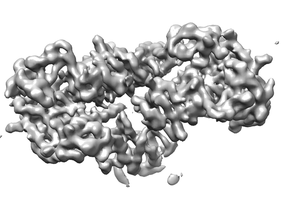
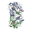
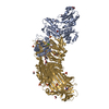
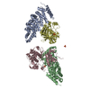
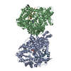
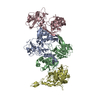
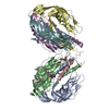
| Title | Insulin Receptor ectodomain in complex with two insulin molecules | ||||||||||||
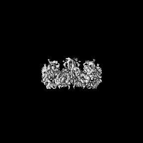
 Map data Map data | Insulin Receptor ectodomain in complex with two insulin molecules | ||||||||||||
 Sample Sample |
| ||||||||||||
 Keywords Keywords | signaling / SIGNALING PROTEIN | ||||||||||||
| Function / homology |  Function and homology information Function and homology informationcellular response to palmitoleic acid / response to L-arginine / regulation of female gonad development / positive regulation of meiotic cell cycle / insulin-like growth factor II binding / positive regulation of developmental growth / male sex determination / insulin receptor complex / insulin-like growth factor I binding / insulin receptor activity ...cellular response to palmitoleic acid / response to L-arginine / regulation of female gonad development / positive regulation of meiotic cell cycle / insulin-like growth factor II binding / positive regulation of developmental growth / male sex determination / insulin receptor complex / insulin-like growth factor I binding / insulin receptor activity / positive regulation of protein-containing complex disassembly / exocrine pancreas development / dendritic spine maintenance / adrenal gland development / insulin binding / cargo receptor activity / negative regulation of glycogen catabolic process / : / negative regulation of fatty acid metabolic process / PTB domain binding / Signaling by Insulin receptor / negative regulation of feeding behavior / IRS activation / Insulin processing / regulation of protein secretion / positive regulation of peptide hormone secretion / neuronal cell body membrane / negative regulation of acute inflammatory response / Regulation of gene expression in beta cells / positive regulation of respiratory burst / alpha-beta T cell activation / amyloid-beta clearance / regulation of embryonic development / insulin receptor substrate binding / positive regulation of receptor internalization / Synthesis, secretion, and deacylation of Ghrelin / epidermis development / negative regulation of protein secretion / positive regulation of dendritic spine maintenance / negative regulation of gluconeogenesis / fatty acid homeostasis / positive regulation of glycogen biosynthetic process / positive regulation of insulin receptor signaling pathway / Signal attenuation / FOXO-mediated transcription of oxidative stress, metabolic and neuronal genes / protein kinase activator activity / negative regulation of lipid catabolic process / negative regulation of respiratory burst involved in inflammatory response / positive regulation of lipid biosynthetic process / negative regulation of oxidative stress-induced intrinsic apoptotic signaling pathway / nitric oxide-cGMP-mediated signaling / regulation of protein localization to plasma membrane / transport across blood-brain barrier / positive regulation of nitric-oxide synthase activity / heart morphogenesis / phosphatidylinositol 3-kinase binding / transport vesicle / Insulin receptor recycling / COPI-mediated anterograde transport / negative regulation of reactive oxygen species biosynthetic process / positive regulation of brown fat cell differentiation / insulin-like growth factor receptor binding / NPAS4 regulates expression of target genes / neuron projection maintenance / positive regulation of mitotic nuclear division / endoplasmic reticulum-Golgi intermediate compartment membrane / Insulin receptor signalling cascade / receptor-mediated endocytosis / dendrite membrane / positive regulation of glycolytic process / endosome lumen / positive regulation of cytokine production / acute-phase response / positive regulation of D-glucose import across plasma membrane / insulin receptor binding / positive regulation of long-term synaptic potentiation / learning / positive regulation of protein secretion / positive regulation of cell differentiation / wound healing / Regulation of insulin secretion / hormone activity / receptor protein-tyrosine kinase / positive regulation of neuron projection development / negative regulation of protein catabolic process / receptor internalization / caveola / regulation of synaptic plasticity / positive regulation of protein localization to nucleus / Golgi lumen / cellular response to growth factor stimulus / male gonad development / cognition / glucose metabolic process / vasodilation / cellular response to insulin stimulus / memory / positive regulation of nitric oxide biosynthetic process / insulin receptor signaling pathway / protein autophosphorylation Similarity search - Function | ||||||||||||
| Biological species |  Homo sapiens (human) / Homo sapiens (human) /  | ||||||||||||
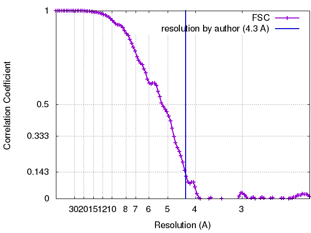
| Method | single particle reconstruction / cryo EM / Resolution: 4.3 Å | ||||||||||||
 Authors Authors | Scapin G / Dandey VP | ||||||||||||
| Funding support |  United States, 3 items United States, 3 items
| ||||||||||||
 Citation Citation | Journal: Nature / Year: 2018 Title: Structure of the insulin receptor-insulin complex by single-particle cryo-EM analysis. Authors: Giovanna Scapin / Venkata P Dandey / Zhening Zhang / Winifred Prosise / Alan Hruza / Theresa Kelly / Todd Mayhood / Corey Strickland / Clinton S Potter / Bridget Carragher / Abstract: The insulin receptor is a dimeric protein that has a crucial role in controlling glucose homeostasis, regulating lipid, protein and carbohydrate metabolism, and modulating brain neurotransmitter ...The insulin receptor is a dimeric protein that has a crucial role in controlling glucose homeostasis, regulating lipid, protein and carbohydrate metabolism, and modulating brain neurotransmitter levels. Insulin receptor dysfunction has been associated with many diseases, including diabetes, cancer and Alzheimer's disease. The primary sequence of the receptor has been known since the 1980s, and is composed of an extracellular portion (the ectodomain, ECD), a single transmembrane helix and an intracellular tyrosine kinase domain. Binding of insulin to the dimeric ECD triggers auto-phosphorylation of the tyrosine kinase domain and subsequent activation of downstream signalling molecules. Biochemical and mutagenesis data have identified two putative insulin-binding sites, S1 and S2. The structures of insulin bound to an ECD fragment containing S1 and of the apo ectodomain have previously been reported, but details of insulin binding to the full receptor and the signal propagation mechanism are still not understood. Here we report single-particle cryo-electron microscopy reconstructions of the 1:2 (4.3 Å) and 1:1 (7.4 Å) complexes of the insulin receptor ECD dimer with insulin. The symmetrical 4.3 Å structure shows two insulin molecules per dimer, each bound between the leucine-rich subdomain L1 of one monomer and the first fibronectin-like domain (FnIII-1) of the other monomer, and making extensive interactions with the α-subunit C-terminal helix (α-CT helix). The 7.4 Å structure has only one similarly bound insulin per receptor dimer. The structures confirm the binding interactions at S1 and define the full S2 binding site. These insulin receptor states suggest that recruitment of the α-CT helix upon binding of the first insulin changes the relative subdomain orientations and triggers downstream signal propagation. | ||||||||||||
| History |
|
- Structure visualization
| Movie |
Movie viewer |
|---|---|
| Structure viewer | EM map: SurfViewMolmilJmol/JSmol |
| Supplemental images |
- Downloads & links
-EMDB archive
| Map data | emd_7462.map.gz | 85.4 MB | EMDB map data format | |
|---|---|---|---|---|
| Header (meta data) | emd-7462-v30.xmlemd-7462.xml | 17.3 KB 17.3 KB | Display Display | EMDB header |
| FSC (resolution estimation) | emd_7462_fsc.xml | 12 KB | Display | FSC data file |
| Images |  emd_7462.png emd_7462.png | 98.6 KB | ||
| Filedesc metadata | emd-7462.cif.gz | 6.4 KB | ||
| Archive directory |  http://ftp.pdbj.org/pub/emdb/structures/EMD-7462ftp://ftp.pdbj.org/pub/emdb/structures/EMD-7462 http://ftp.pdbj.org/pub/emdb/structures/EMD-7462ftp://ftp.pdbj.org/pub/emdb/structures/EMD-7462 | HTTPS FTP |
-Related structure data
| Related structure data |  6ce9MC  7461C  7463C  6ce7C  6cebC C: citing same article ( M: atomic model generated by this map |
|---|---|
| Similar structure data |


-Links
| EMDB pages | EMDB (EBI/PDBe) / EMDataResource |
|---|---|
| Related items in Molecule of the Month |


-Map
| File | Download / File: emd_7462.map.gz / Format: CCP4 / Size: 91.1 MB / Type: IMAGE STORED AS FLOATING POINT NUMBER (4 BYTES) | ||||||||||||||||||||||||||||||||||||||||||||||||||||||||||||
|---|---|---|---|---|---|---|---|---|---|---|---|---|---|---|---|---|---|---|---|---|---|---|---|---|---|---|---|---|---|---|---|---|---|---|---|---|---|---|---|---|---|---|---|---|---|---|---|---|---|---|---|---|---|---|---|---|---|---|---|---|---|
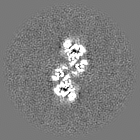
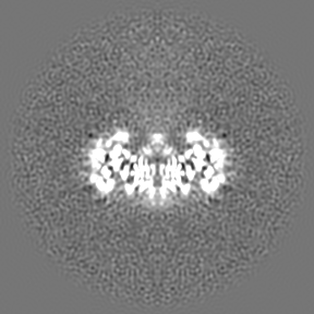
| Annotation | Insulin Receptor ectodomain in complex with two insulin molecules | ||||||||||||||||||||||||||||||||||||||||||||||||||||||||||||
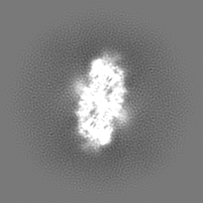
| Projections & slices | Image control
Images are generated by Spider. | ||||||||||||||||||||||||||||||||||||||||||||||||||||||||||||
| Voxel size | X=Y=Z: 1.1 Å | ||||||||||||||||||||||||||||||||||||||||||||||||||||||||||||
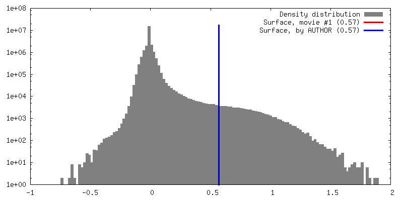
| Density |
| ||||||||||||||||||||||||||||||||||||||||||||||||||||||||||||
| Symmetry | Space group: 1 | ||||||||||||||||||||||||||||||||||||||||||||||||||||||||||||
| Details | EMDB XML:
CCP4 map header:
| ||||||||||||||||||||||||||||||||||||||||||||||||||||||||||||
 Z (Sec.)
 Y (Row.)
 X (Col.)


-Supplemental data
- Sample components
+Entire : Insulin Receptor Ectodomain in complex with two insulin molecules
+Supramolecule #1: Insulin Receptor Ectodomain in complex with two insulin molecules
+Supramolecule #2: Insulin receptor
+Supramolecule #3: Insulin receptor subunit alpha
+Supramolecule #4: Insulin A chain
+Supramolecule #5: Insulin B chain
+Macromolecule #1: Insulin receptor
+Macromolecule #2: Insulin receptor
+Macromolecule #3: Insulin A chain
+Macromolecule #4: Insulin B chain
+Macromolecule #9: 2-acetamido-2-deoxy-beta-D-glucopyranose

-Experimental details
-Structure determination
| Method | cryo EM |
|---|---|
 Processing Processing | single particle reconstruction |
| Aggregation state | particle |
-Sample preparation
| Concentration | 0.3 mg/mL |
|---|---|
| Buffer | pH: 7.5 / Details: Hepes Saline (HBS) |
| Vitrification | Cryogen name: ETHANE / Chamber humidity: 80 % / Chamber temperature: 293 K / Instrument: HOMEMADE PLUNGER / Details: Grids made with SpotItOn. |
- Electron microscopy
| Microscope | FEI TITAN KRIOS |
|---|---|
| Image recording | Film or detector model: GATAN K2 QUANTUM (4k x 4k) / Detector mode: COUNTING / Average electron dose: 45.5 e/Å2 |
| Electron beam | Acceleration voltage: 300 kV / Electron source:  FIELD EMISSION GUN FIELD EMISSION GUN |
| Electron optics | Illumination mode: OTHER / Imaging mode: BRIGHT FIELD |
| Experimental equipment |  Model: Titan Krios / Image courtesy: FEI Company |
+Image processing
-Atomic model buiding 1
| Refinement | Protocol: RIGID BODY FIT |
|---|---|
| Output model | PDB-6ce9: |
