 Movie
Movie Controller

[English] 日本語
 Yorodumi
- PDB-1slq: Crystal structure of the trimeric state of the rhesus rotavirus V... -
+ Open data

- Basic information
| Entry | Database: PDB / ID: 1slq | ||||||
|---|---|---|---|---|---|---|---|
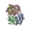
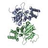
| Title | Crystal structure of the trimeric state of the rhesus rotavirus VP4 membrane interaction domain, VP5CT | ||||||
 Components Components | VP4 | ||||||
 Keywords Keywords | VIRAL PROTEIN / beta sandwich / greek key / alpha helical triple coiled-coil / membrane penetration protein / non-enveloped virus / spike protein | ||||||
| Function / homology |  Function and homology information Function and homology informationhost cell rough endoplasmic reticulum / permeabilization of host organelle membrane involved in viral entry into host cell / host cytoskeleton / viral outer capsid / host cell endoplasmic reticulum-Golgi intermediate compartment / virion attachment to host cell / host cell plasma membrane Similarity search - Function | ||||||
| Biological species |  Rhesus rotavirus Rhesus rotavirus | ||||||
| Method |  X-RAY DIFFRACTION / SYNCHROTRON / MAD / Resolution: 3.2 Å X-RAY DIFFRACTION / SYNCHROTRON / MAD / Resolution: 3.2 Å | ||||||
 Authors Authors | Dormitzer, P.R. / Nason, E.B. / Prasad, B.V.V. / Harrison, S.C. | ||||||
 Citation Citation | Journal: Nature / Year: 2004 Title: Structural rearrangements in the membrane penetration protein of a non-enveloped virus. Authors: Dormitzer, P.R. / Nason, E.B. / Prasad, B.V. / Harrison, S.C. #1: Journal: J.Virol. / Year: 2001Title: Proteolysis of monomeric recombinant rotavirus VP4 yields an oligomeric VP5* core Authors: Dormitzer, P.R. / Greenberg, H.B. / Harrison, S.C. #2: Journal: Embo J. / Year: 2002Title: The rhesus rotavirus VP4 sialic acid binding domain has a galectin fold with a novel carbohydrate binding site Authors: Dormitzer, P.R. / Sun, Z.-Y.J. / Wagner, G. / Harrison, S.C. #3: Journal: Acta Crystallogr.,Sect.D / Year: 2003Title: A statistic for local intensity differences: robustness to anisotropy and pseudo-centering and utility for detecting twinning Authors: Padilla, J.E. / Yeates, T.O. | ||||||
| History |
|
- Structure visualization
| Structure viewer | Molecule: MolmilJmol/JSmol |
|---|
- Downloads & links
-Download
| PDBx/mmCIF format | 1slq.cif.gz | 291.7 KB | Display | PDBx/mmCIF format |
|---|---|---|---|---|
| PDB format | pdb1slq.ent.gz | 244 KB | Display | PDB format |
| PDBx/mmJSON format | 1slq.json.gz | Tree view | PDBx/mmJSON format | |
| Others |  Other downloads Other downloads |
-Validation report
| Arichive directory | https://data.pdbj.org/pub/pdb/validation_reports/sl/1slqftp://data.pdbj.org/pub/pdb/validation_reports/sl/1slq | HTTPS FTP |
|---|
-Related structure data
| Related structure data | |
|---|---|
| Similar structure data |


-Links
 PDBj
PDBj- Assembly
| Deposited unit | 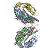
| ||||||||
|---|---|---|---|---|---|---|---|---|---|
| 1 | 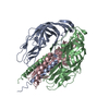
| ||||||||
| 2 | 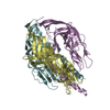
| ||||||||
| 3 | 
| ||||||||
| 4 | 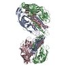
| ||||||||
| Unit cell |
| ||||||||
| Details | The biological unit is a homotrimer. Each asymmetric unit in space group P4(2)22 contains 2 trimers. |
-Components
| #1: Protein | Mass: 31307.945 Da / Num. of mol.: 6 Fragment: Trimeric conformation of the membrane interaction domain, VP5CT Source method: isolated from a genetically manipulated source Source: (gene. exp.) Rhesus rotavirus / Species: Rotavirus A / Gene: gene segment 4 / Production host:   Spodoptera frugiperda (fall armyworm) / Strain (production host): Sf9 / References: UniProt: Q91HI9, UniProt: P12473*PLUS Spodoptera frugiperda (fall armyworm) / Strain (production host): Sf9 / References: UniProt: Q91HI9, UniProt: P12473*PLUS |
|---|
-Experimental details
-Experiment
| Experiment | Method: X-RAY DIFFRACTION / Number of used crystals: 2 |
|---|
- Sample preparation
| Crystal | Density Matthews: 4.18 Å3/Da / Density % sol: 70.56 % | |||||||||||||||||||||||||||||||||||||||||||||
|---|---|---|---|---|---|---|---|---|---|---|---|---|---|---|---|---|---|---|---|---|---|---|---|---|---|---|---|---|---|---|---|---|---|---|---|---|---|---|---|---|---|---|---|---|---|---|
| Crystal grow | Temperature: 296 K / Method: vapor diffusion, hanging drop / pH: 6.9 Details: ammonium sulfate, MPD, Pipes, Tris, EDTA, sodium azide, benzamidine, pH 6.9, VAPOR DIFFUSION, HANGING DROP, temperature 296K | |||||||||||||||||||||||||||||||||||||||||||||
| Crystal grow | *PLUS Temperature: 23 ℃ / pH: 8 | |||||||||||||||||||||||||||||||||||||||||||||
| Components of the solutions | *PLUS
|
-Data collection
| Diffraction |
| ||||||||||||||||||
|---|---|---|---|---|---|---|---|---|---|---|---|---|---|---|---|---|---|---|---|
| Diffraction source |
| ||||||||||||||||||
| Detector |
| ||||||||||||||||||
| Radiation |
| ||||||||||||||||||
| Radiation wavelength | Wavelength: 0.916 Å / Relative weight: 1 | ||||||||||||||||||
| Reflection | Resolution: 3.2→20 Å / Num. all: 51973 / Num. obs: 51973 / % possible obs: 99.8 % / Redundancy: 8.4 % / Rmerge(I) obs: 0.057 / Rsym value: 0.057 / Net I/σ(I): 23.6 | ||||||||||||||||||
| Reflection shell | Resolution: 3.2→3.27 Å / Redundancy: 7.1 % / Rmerge(I) obs: 0.12 / Mean I/σ(I) obs: 8.8 / Num. unique all: 3446 / Rsym value: 0.12 / % possible all: 99.8 | ||||||||||||||||||
| Reflection | *PLUS | ||||||||||||||||||
| Reflection shell | *PLUS % possible obs: 99.8 % / Num. unique obs: 3446 / Rmerge(I) obs: 0.12 |

- Processing
| Software |
| |||||||||||||||||||||||||
|---|---|---|---|---|---|---|---|---|---|---|---|---|---|---|---|---|---|---|---|---|---|---|---|---|---|---|
| Refinement | Method to determine structure: MAD / Resolution: 3.2→20 Å / Isotropic thermal model: isotropic by group / Cross valid method: THROUGHOUT / σ(F): 0.01 / Stereochemistry target values: Engh & Huber Details: An initial model was built to maps calculated with MAD experimental data. The MAD data were collected from crystals of selenomethionine-substituted VP5CT that formed using ethanol as a ...Details: An initial model was built to maps calculated with MAD experimental data. The MAD data were collected from crystals of selenomethionine-substituted VP5CT that formed using ethanol as a precipitant. Refinement proceeded with starting phases from this model and amplitudes from higher resolution data collected from native VP5CT crystals that formed using ammonium sulfate with MPD as a precipitant. All stages of model building and refinement took advantage of 6-fold non-crystallographic symmetry. The resolution of the model is limited by perfect hemihedral twinning, which becomes significant at higher resolutions.
| |||||||||||||||||||||||||
| Displacement parameters | Biso mean: 91.45 Å2 | |||||||||||||||||||||||||
| Refine analyze |
| |||||||||||||||||||||||||
| Refinement step | Cycle: LAST / Resolution: 3.2→20 Å
| |||||||||||||||||||||||||
| Refine LS restraints |
| |||||||||||||||||||||||||
| LS refinement shell | Resolution: 3.2→3.27 Å
| |||||||||||||||||||||||||
| Software | *PLUS Name: CNS / Version: 1 / Classification: refinement | |||||||||||||||||||||||||
| Refinement | *PLUS Highest resolution: 3.2 Å / Lowest resolution: 20 Å / σ(F): 0.01 / Rfactor obs: 0.308 | |||||||||||||||||||||||||
| Solvent computation | *PLUS | |||||||||||||||||||||||||
| Displacement parameters | *PLUS | |||||||||||||||||||||||||
| Refine LS restraints | *PLUS
| |||||||||||||||||||||||||
| LS refinement shell | *PLUS Rfactor Rfree: 0.488 / Rfactor Rwork: 0.443 |
