 Movie
Movie Controller

[English] 日本語
 Yorodumi
- PDB-1kri: NMR Solution Structures of the Rhesus Rotavirus VP4 Sialic Acid B... -
+ Open data

- Basic information
| Entry | Database: PDB / ID: 1kri | ||||||
|---|---|---|---|---|---|---|---|
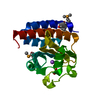
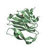
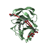
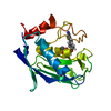
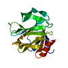
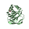
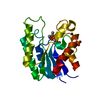
| Title | NMR Solution Structures of the Rhesus Rotavirus VP4 Sialic Acid Binding Domain without Ligand | ||||||
 Components Components | VP4 | ||||||
 Keywords Keywords | VIRAL PROTEIN / rotavirus / VP4 / VP8* / spike protein / outer capsid / sialic acid / hemagglutinin / cell attachment / neutralization antigen / lectin / galectin fold | ||||||
| Function / homology |  Function and homology information Function and homology informationhost cell rough endoplasmic reticulum / permeabilization of host organelle membrane involved in viral entry into host cell / host cytoskeleton / viral outer capsid / host cell endoplasmic reticulum-Golgi intermediate compartment / virion attachment to host cell / host cell plasma membrane Similarity search - Function | ||||||
| Biological species |  Rhesus rotavirus Rhesus rotavirus | ||||||
| Method | SOLUTION NMR / simulated annealing | ||||||
 Authors Authors | Dormitzer, P.R. / Sun, Z.-Y.J. / Wagner, G. / Harrison, S.C. | ||||||
 Citation Citation | Journal: Embo J. / Year: 2002 Title: The Rhesus Rotavirus VP4 Sialic Acid Binding Domain has a Galectin Fold with a Novel Carbohydrate Binding Site Authors: Dormitzer, P.R. / Sun, Z.-Y.J. / Wagner, G. / Harrison, S.C. #1: Journal: J.Virol. / Year: 2001Title: Proteolysis of Monomeric Recombinant Rotavirus VP4 Yields an Oligomeric VP5* Core Authors: Dormitzer, P.R. / Greenberg, H.B. / Harrison, S.C. | ||||||
| History |
| ||||||
| Remark 700 | sheet determination method: author |
- Structure visualization
| Structure viewer | Molecule: MolmilJmol/JSmol |
|---|
- Downloads & links
-Download
| PDBx/mmCIF format | 1kri.cif.gz | 961.7 KB | Display | PDBx/mmCIF format |
|---|---|---|---|---|
| PDB format | pdb1kri.ent.gz | 802.6 KB | Display | PDB format |
| PDBx/mmJSON format | 1kri.json.gz | Tree view | PDBx/mmJSON format | |
| Others |  Other downloads Other downloads |
-Validation report
| Arichive directory | https://data.pdbj.org/pub/pdb/validation_reports/kr/1kriftp://data.pdbj.org/pub/pdb/validation_reports/kr/1kri | HTTPS FTP |
|---|
-Related structure data


-Links
 PDBj
PDBj
- Assembly
| Deposited unit | 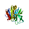
| |||||||||
|---|---|---|---|---|---|---|---|---|---|---|
| 1 |
| |||||||||
| NMR ensembles |
|
-Components
| #1: Protein | Mass: 20816.887 Da / Num. of mol.: 1 / Fragment: sialic acid binding domain (residues 46-231) Source method: isolated from a genetically manipulated source Source: (gene. exp.) Rhesus rotavirus / Species: Rotavirus A / Gene: segment 4 / Plasmid: pGEX-VP8(46-231) / Production host:  |
|---|
-Experimental details
-Experiment
| Experiment | Method: SOLUTION NMR | ||||||||||||||||||||||||||||||||||||||||||||||||||||||||||||||||||||||||||||
|---|---|---|---|---|---|---|---|---|---|---|---|---|---|---|---|---|---|---|---|---|---|---|---|---|---|---|---|---|---|---|---|---|---|---|---|---|---|---|---|---|---|---|---|---|---|---|---|---|---|---|---|---|---|---|---|---|---|---|---|---|---|---|---|---|---|---|---|---|---|---|---|---|---|---|---|---|---|
| NMR experiment |
| ||||||||||||||||||||||||||||||||||||||||||||||||||||||||||||||||||||||||||||
| NMR details | Text: The N-terminal 19 residues (A46 to V64 of VP4) and the C-terminal 7 residues (P225 to R231 of VP4) are disordered and are not included in the models. |
 HSQC
HSQC- Sample preparation
| Details |
| |||||||||||||||||||||
|---|---|---|---|---|---|---|---|---|---|---|---|---|---|---|---|---|---|---|---|---|---|---|
| Sample conditions | Ionic strength: 20 mM NaPO4, 10 mM NaCl, 0.02% Na azide / pH: 7 / Pressure: ambient / Temperature: 298 K | |||||||||||||||||||||
| Crystal grow | *PLUS Method: other / Details: NMR |
-NMR measurement
| NMR spectrometer |
|
|---|
- Processing
| NMR software |
| ||||||||||||||||||||||||||||
|---|---|---|---|---|---|---|---|---|---|---|---|---|---|---|---|---|---|---|---|---|---|---|---|---|---|---|---|---|---|
| Refinement | Method: simulated annealing / Software ordinal: 1 Details: The models are based on a total of 1993 constraints (12.5 constraints per residue): 1793 NOE-derived distance constraints; 116 TALOS-derived dihedral angle constraints; and 84 hydrogen bond ...Details: The models are based on a total of 1993 constraints (12.5 constraints per residue): 1793 NOE-derived distance constraints; 116 TALOS-derived dihedral angle constraints; and 84 hydrogen bond constraints, based on the identification of slow exchange amide protons in D2O NOESY and TOCSY experiments, on characteristic NOE patterns for alpha-helices and beta-sheets, and on the proximity and orientation of potential hydrogen bond partners in annealed structures. The final set of 20 structures contains no violations of NOE distance constraints greater than 0.15 angstroms and no violations of dihedral angle constraints greater than 5 degrees. | ||||||||||||||||||||||||||||
| NMR representative | Selection criteria: least restraint violations | ||||||||||||||||||||||||||||
| NMR ensemble | Conformer selection criteria: structures with the least restraint violations Conformers calculated total number: 25 / Conformers submitted total number: 20 |
