 Movie
Movie Controller

[English] 日本語
 Yorodumi
- PDB-5ymu: Functional and structural characterization of P[19] rotavirus VP8... -
+ Open data

- Basic information
| Entry | Database: PDB / ID: 5ymu | ||||||
|---|---|---|---|---|---|---|---|
| Title | Functional and structural characterization of P[19] rotavirus VP8* interaction with histo-blood group antigens | ||||||
 Components Components | Outer capsid protein VP4 | ||||||
 Keywords Keywords | VIRAL PROTEIN / Rotavirus / P[19] VP8* | ||||||
| Function / homology |  Function and homology information Function and homology informationhost cell rough endoplasmic reticulum / permeabilization of host organelle membrane involved in viral entry into host cell / host cytoskeleton / viral outer capsid / host cell endoplasmic reticulum-Golgi intermediate compartment / virion attachment to host cell / host cell plasma membrane / membrane Similarity search - Function | ||||||
| Biological species |  Porcine rotavirus A Porcine rotavirus A | ||||||
| Method |  X-RAY DIFFRACTION / SYNCHROTRON / MOLECULAR REPLACEMENT / Resolution: 1.8 Å X-RAY DIFFRACTION / SYNCHROTRON / MOLECULAR REPLACEMENT / Resolution: 1.8 Å | ||||||
 Authors Authors | Sun, X. / Duan, Z. | ||||||
| Funding support |  China, 1items China, 1items
| ||||||
 Citation Citation | Journal: J. Virol. / Year: 2018 Title: Glycan Binding Specificity and Mechanism of Human and Porcine P[6]/P[19] Rotavirus VP8*s. Authors: Sun, X. / Li, D. / Qi, J. / Chai, W. / Wang, L. / Wang, L. / Peng, R. / Wang, H. / Zhang, Q. / Pang, L. / Kong, X. / Wang, H. / Jin, M. / Gao, G.F. / Duan, Z. | ||||||
| History |
|
- Structure visualization
| Structure viewer | Molecule: MolmilJmol/JSmol |
|---|
- Downloads & links
-Download
| PDBx/mmCIF format | 5ymu.cif.gz | 292.4 KB | Display | PDBx/mmCIF format |
|---|---|---|---|---|
| PDB format | pdb5ymu.ent.gz | 239.9 KB | Display | PDB format |
| PDBx/mmJSON format | 5ymu.json.gz | Tree view | PDBx/mmJSON format | |
| Others |  Other downloads Other downloads |
-Validation report
| Arichive directory | https://data.pdbj.org/pub/pdb/validation_reports/ym/5ymuftp://data.pdbj.org/pub/pdb/validation_reports/ym/5ymu | HTTPS FTP |
|---|
-Related structure data
| Related structure data |  5ymsC  5ymtC  5gj6S S: Starting model for refinement C: citing same article ( |
|---|---|
| Similar structure data |


-Links
 PDBj
PDBj- Assembly
| Deposited unit | 
| ||||||||
|---|---|---|---|---|---|---|---|---|---|
| 1 | 
| ||||||||
| 2 | 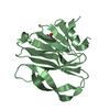
| ||||||||
| 3 | 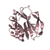
| ||||||||
| 4 | 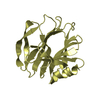
| ||||||||
| Unit cell |
| ||||||||
| Components on special symmetry positions |
|
-Components
| #1: Protein | Mass: 18513.578 Da / Num. of mol.: 4 Source method: isolated from a genetically manipulated source Source: (gene. exp.) Porcine rotavirus AProduction host: References: UniProt: A0A2U8JDD5*PLUS #2: Chemical |   Mass: 92.094 Da / Num. of mol.: 3 / Source method: obtained synthetically / Formula: C3H8O3 Mass: 92.094 Da / Num. of mol.: 3 / Source method: obtained synthetically / Formula: C3H8O3#3: Water | ChemComp-HOH / |  Mass: 18.015 Da / Num. of mol.: 692 / Source method: isolated from a natural source / Formula: H2O Mass: 18.015 Da / Num. of mol.: 692 / Source method: isolated from a natural source / Formula: H2O |
|---|
-Experimental details
-Experiment
| Experiment | Method: X-RAY DIFFRACTION / Number of used crystals: 1 |
|---|
- Sample preparation
| Crystal | Density Matthews: 2.77 Å3/Da / Density % sol: 55.64 % |
|---|---|
| Crystal grow | Temperature: 291 K / Method: vapor diffusion, sitting drop / pH: 6 Details: 0.1 M MES monohydrate pH 6.0, 25% w/v polyethylene glycol 4000 |
-Data collection
| Diffraction | Mean temperature: 123 K |
|---|---|
| Diffraction source | Source: SYNCHROTRON / Site: SSRF / Beamline: BL17U1 / Wavelength: 0.97775 Å |
| Detector | Type: ADSC QUANTUM 315r / Detector: CCD / Date: Jul 3, 2017 |
| Radiation | Protocol: SINGLE WAVELENGTH / Monochromatic (M) / Laue (L): M / Scattering type: x-ray |
| Radiation wavelength | Wavelength: 0.97775 Å / Relative weight: 1 |
| Reflection | Resolution: 1.796→50 Å / Num. obs: 74097 / % possible obs: 100 % / Redundancy: 10.5 % / Net I/σ(I): 21.895 |
- Processing
| Software |
| ||||||||||||||||||||||||||||||||||||||||
|---|---|---|---|---|---|---|---|---|---|---|---|---|---|---|---|---|---|---|---|---|---|---|---|---|---|---|---|---|---|---|---|---|---|---|---|---|---|---|---|---|---|
| Refinement | Method to determine structure: MOLECULAR REPLACEMENT Starting model: 5GJ6 Resolution: 1.8→41.86 Å / SU ML: 0.17 / Cross valid method: FREE R-VALUE / σ(F): 1.96 / Phase error: 19.03
| ||||||||||||||||||||||||||||||||||||||||
| Solvent computation | Shrinkage radii: 0.9 Å / VDW probe radii: 1.11 Å | ||||||||||||||||||||||||||||||||||||||||
| Displacement parameters | Biso mean: 24.06 Å2 | ||||||||||||||||||||||||||||||||||||||||
| Refinement step | Cycle: LAST / Resolution: 1.8→41.86 Å
| ||||||||||||||||||||||||||||||||||||||||
| Refinement TLS params. | Method: refined / Origin x: 36.3251 Å / Origin y: -37.9375 Å / Origin z: 25.8261 Å
| ||||||||||||||||||||||||||||||||||||||||
| Refinement TLS group | Selection details: ALL |
