 Movie
Movie Controller


+ Open data

- Basic information
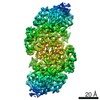
| Entry | Database: PDB / ID: 6oai | |||||||||
|---|---|---|---|---|---|---|---|---|---|---|
| Title | Crystal structure of P[6] rotavirus vp8* complexed with LNFPI | |||||||||
 Components Components | Protease-sensitive outer capsid protein | |||||||||
 Keywords Keywords | VIRUS / Rotavirus / host receptor interaction | |||||||||
| Function / homology |  Function and homology information Function and homology information | |||||||||
| Biological species |  Human rotavirus A Human rotavirus A | |||||||||
| Method |  X-RAY DIFFRACTION / SYNCHROTRON / MOLECULAR REPLACEMENT / Resolution: 1.9 Å X-RAY DIFFRACTION / SYNCHROTRON / MOLECULAR REPLACEMENT / Resolution: 1.9 Å | |||||||||
 Authors Authors | Xu, S. / Liu, Y. / Jiang, X. / Kennedy, M.A. | |||||||||
 Citation Citation | Journal: Plos Pathog. / Year: 2020 Title: Molecular basis of P[II] major human rotavirus VP8* domain recognition of histo-blood group antigens. Authors: Xu, S. / Ahmed, L.U. / Stuckert, M.R. / McGinnis, K.R. / Liu, Y. / Tan, M. / Huang, P. / Zhong, W. / Zhao, D. / Jiang, X. / Kennedy, M.A. | |||||||||
| History |
|
- Structure visualization
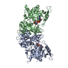
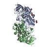
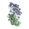
| Structure viewer | Molecule: MolmilJmol/JSmol |
|---|
- Downloads & links
-Download
| PDBx/mmCIF format | 6oai.cif.gz | 145 KB | Display | PDBx/mmCIF format |
|---|---|---|---|---|
| PDB format | pdb6oai.ent.gz | 111.8 KB | Display | PDB format |
| PDBx/mmJSON format | 6oai.json.gz | Tree view | PDBx/mmJSON format | |
| Others |  Other downloads Other downloads |
-Validation report
| Arichive directory | https://data.pdbj.org/pub/pdb/validation_reports/oa/6oaiftp://data.pdbj.org/pub/pdb/validation_reports/oa/6oai | HTTPS FTP |
|---|
-Related structure data
| Related structure data |  6niwSC S: Starting model for refinement C: citing same article ( |
|---|---|
| Similar structure data |


-Links
 PDBj
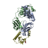
PDBj- Assembly
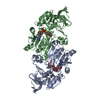
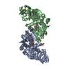
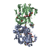
| Deposited unit | 
| ||||||||
|---|---|---|---|---|---|---|---|---|---|
| 1 |
| ||||||||
| 2 | 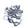
| ||||||||
| 3 | 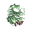
| ||||||||
| 4 | 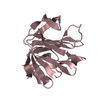
| ||||||||
| 5 | 
| ||||||||
| Unit cell |
|
-Components
| #1: Protein | Mass: 18443.498 Da / Num. of mol.: 4 / Fragment: VP8* domain, residues 49-207 Source method: isolated from a genetically manipulated source Source: (gene. exp.) Human rotavirus A / Production host:  #2: Polysaccharide | alpha-L-fucopyranose-(1-2)-beta-D-galactopyranose-(1-3)-2-acetamido-2-deoxy-beta-D-glucopyranose-(1- ...alpha-L-fucopyranose-(1-2)-beta-D-galactopyranose-(1-3)-2-acetamido-2-deoxy-beta-D-glucopyranose-(1-3)-beta-D-galactopyranose-(1-4)-beta-D-glucopyranose | Source method: isolated from a genetically manipulated source #3: Water | ChemComp-HOH / |  Mass: 18.015 Da / Num. of mol.: 148 / Source method: isolated from a natural source / Formula: H2O Mass: 18.015 Da / Num. of mol.: 148 / Source method: isolated from a natural source / Formula: H2O |
|---|
-Experimental details
-Experiment
| Experiment | Method: X-RAY DIFFRACTION / Number of used crystals: 1 |
|---|
- Sample preparation
| Crystal | Density Matthews: 2.18 Å3/Da / Density % sol: 43.66 % |
|---|---|
| Crystal grow | Temperature: 293 K / Method: vapor diffusion, hanging drop / pH: 5.6 Details: 0.5 M Ammonium sulfate, 0.1 M Sodium citrate tribasic dihydrate, 1.0 M Lithium sulfate monohydrate |
-Data collection
| Diffraction | Mean temperature: 100 K / Serial crystal experiment: N | ||||||||||||||||||||||||
|---|---|---|---|---|---|---|---|---|---|---|---|---|---|---|---|---|---|---|---|---|---|---|---|---|---|
| Diffraction source | Source: SYNCHROTRON / Site: APS  / Beamline: 31-ID / Wavelength: 0.97931 Å / Beamline: 31-ID / Wavelength: 0.97931 Å | ||||||||||||||||||||||||
| Detector | Type: DECTRIS PILATUS3 S 6M / Detector: PIXEL / Date: Mar 1, 2019 | ||||||||||||||||||||||||
| Radiation | Protocol: SINGLE WAVELENGTH / Monochromatic (M) / Laue (L): M / Scattering type: x-ray | ||||||||||||||||||||||||
| Radiation wavelength | Wavelength: 0.97931 Å / Relative weight: 1 | ||||||||||||||||||||||||
| Reflection | Resolution: 1.9→74.89 Å / Num. obs: 48808 / % possible obs: 97.3 % / Redundancy: 3.4 % / CC1/2: 0.998 / Rmerge(I) obs: 0.073 / Rpim(I) all: 0.046 / Rrim(I) all: 0.087 / Net I/σ(I): 12.4 / Num. measured all: 164606 | ||||||||||||||||||||||||
| Reflection shell | Diffraction-ID: 1
|
- Processing
| Software |
| ||||||||||||||||||||||||||||||||||||||||||||||||||||||||||||
|---|---|---|---|---|---|---|---|---|---|---|---|---|---|---|---|---|---|---|---|---|---|---|---|---|---|---|---|---|---|---|---|---|---|---|---|---|---|---|---|---|---|---|---|---|---|---|---|---|---|---|---|---|---|---|---|---|---|---|---|---|---|
| Refinement | Method to determine structure: MOLECULAR REPLACEMENT Starting model: 6NIW Resolution: 1.9→45.95 Å / Cor.coef. Fo:Fc: 0.947 / Cor.coef. Fo:Fc free: 0.914 / SU B: 4.182 / SU ML: 0.12 / Cross valid method: THROUGHOUT / σ(F): 0 / ESU R: 0.177 / ESU R Free: 0.166 Details: HYDROGENS HAVE BEEN ADDED IN THE RIDING POSITIONS U VALUES : REFINED INDIVIDUALLY
| ||||||||||||||||||||||||||||||||||||||||||||||||||||||||||||
| Solvent computation | Ion probe radii: 0.8 Å / Shrinkage radii: 0.8 Å / VDW probe radii: 1.2 Å | ||||||||||||||||||||||||||||||||||||||||||||||||||||||||||||
| Displacement parameters | Biso max: 77.03 Å2 / Biso mean: 19.793 Å2 / Biso min: 4.2 Å2
| ||||||||||||||||||||||||||||||||||||||||||||||||||||||||||||
| Refinement step | Cycle: final / Resolution: 1.9→45.95 Å
| ||||||||||||||||||||||||||||||||||||||||||||||||||||||||||||
| Refine LS restraints |
| ||||||||||||||||||||||||||||||||||||||||||||||||||||||||||||
| LS refinement shell | Resolution: 1.898→1.948 Å / Rfactor Rfree error: 0 / Total num. of bins used: 20
|
