 Movie
Movie Controller


+ Open data

- Basic information
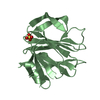
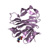
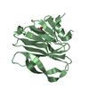
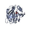
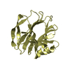
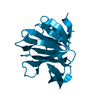
| Entry | Database: PDB / ID: 5caz | ||||||
|---|---|---|---|---|---|---|---|
| Title | Crystallographic structure of apo human rotavirus K8 VP8* | ||||||
 Components Components | Outer capsid protein VP4 | ||||||
 Keywords Keywords | viral protein / sugard binding protein / carbohydrate-recognizing protein / lectin / rotavirus / VP8* | ||||||
| Function / homology |  Function and homology information Function and homology informationhost cell rough endoplasmic reticulum / permeabilization of host organelle membrane involved in viral entry into host cell / host cytoskeleton / viral outer capsid / host cell endoplasmic reticulum-Golgi intermediate compartment / virion attachment to host cell / host cell plasma membrane Similarity search - Function | ||||||
| Biological species |  Rotavirus A Rotavirus A | ||||||
| Method |  X-RAY DIFFRACTION / SYNCHROTRON / MOLECULAR REPLACEMENT / Resolution: 1.8 Å X-RAY DIFFRACTION / SYNCHROTRON / MOLECULAR REPLACEMENT / Resolution: 1.8 Å | ||||||
 Authors Authors | Yu, X. / Blanchard, H. | ||||||
| Funding support |  Australia, 1items Australia, 1items
| ||||||
 Citation Citation | Journal: Chembiochem / Year: 2015 Title: Substantial Receptor-induced Structural Rearrangement of Rotavirus VP8*: Potential Implications for Cross-Species Infection. Authors: Yu, X. / Mishra, R. / Holloway, G. / von Itzstein, M. / Coulson, B.S. / Blanchard, H. | ||||||
| History |
|
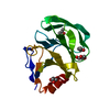
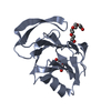
- Structure visualization
| Structure viewer | Molecule: MolmilJmol/JSmol |
|---|
- Downloads & links
-Download
| PDBx/mmCIF format | 5caz.cif.gz | 53.7 KB | Display | PDBx/mmCIF format |
|---|---|---|---|---|
| PDB format | pdb5caz.ent.gz | 36.4 KB | Display | PDB format |
| PDBx/mmJSON format | 5caz.json.gz | Tree view | PDBx/mmJSON format | |
| Others |  Other downloads Other downloads |
-Validation report
| Arichive directory | https://data.pdbj.org/pub/pdb/validation_reports/ca/5cazftp://data.pdbj.org/pub/pdb/validation_reports/ca/5caz | HTTPS FTP |
|---|
-Related structure data
| Related structure data | 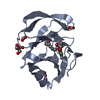 5ca6C 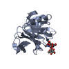 5cb7C 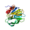 4drrS C: citing same article ( S: Starting model for refinement |
|---|---|
| Similar structure data |

-Links
 PDBj
PDBj
- Assembly
| Deposited unit | 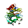
| ||||||||
|---|---|---|---|---|---|---|---|---|---|
| 1 |
| ||||||||
| Unit cell |
|
-Components
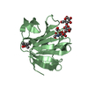
| #1: Protein | Mass: 18379.334 Da / Num. of mol.: 1 / Fragment: unp residues 64-224 / Mutation: Y149D Source method: isolated from a genetically manipulated source Source: (gene. exp.) Rotavirus A (strain Human/Japan/K8/1977 G1-P3A[9]-Ix-Rx-Cx-Mx-A1-Nx-Tx-Ex-H3)Strain: Human/Japan/K8/1977 G1-P3A[9]-Ix-Rx-Cx-Mx-A1-Nx-Tx-Ex-H3 Production host:  | ||||||
|---|---|---|---|---|---|---|---|
| #2: Chemical | ChemComp-IPA /   Mass: 60.095 Da / Num. of mol.: 7 / Source method: obtained synthetically / Formula: C3H8O Mass: 60.095 Da / Num. of mol.: 7 / Source method: obtained synthetically / Formula: C3H8O#3: Chemical | ChemComp-SO4 / |   Mass: 96.063 Da / Num. of mol.: 1 / Source method: obtained synthetically / Formula: SO4 Mass: 96.063 Da / Num. of mol.: 1 / Source method: obtained synthetically / Formula: SO4#4: Chemical | ChemComp-GOL / |   Mass: 92.094 Da / Num. of mol.: 1 / Source method: obtained synthetically / Formula: C3H8O3 Mass: 92.094 Da / Num. of mol.: 1 / Source method: obtained synthetically / Formula: C3H8O3#5: Water | ChemComp-HOH / |  Mass: 18.015 Da / Num. of mol.: 183 / Source method: isolated from a natural source / Formula: H2O Mass: 18.015 Da / Num. of mol.: 183 / Source method: isolated from a natural source / Formula: H2O |
-Experimental details
-Experiment
| Experiment | Method: X-RAY DIFFRACTION |
|---|
- Sample preparation
| Crystal | Density Matthews: 2.31 Å3/Da / Density % sol: 46.81 % |
|---|---|
| Crystal grow | Temperature: 293 K / Method: vapor diffusion, hanging drop / Details: 2 M ammonium sulphate and 5% (v/v) isopropanol |
-Data collection
| Diffraction | Mean temperature: 100 K |
|---|---|
| Diffraction source | Source: SYNCHROTRON / Site: Australian Synchrotron / Beamline: MX2 / Wavelength: 0.954 Å |
| Detector | Type: ADSC QUANTUM 315r / Detector: CCD / Date: Nov 27, 2012 |
| Radiation | Protocol: SINGLE WAVELENGTH / Monochromatic (M) / Laue (L): M / Scattering type: x-ray |
| Radiation wavelength | Wavelength: 0.954 Å / Relative weight: 1 |
| Reflection | Resolution: 1.8→54.4 Å / Num. obs: 15568 / % possible obs: 100 % / Redundancy: 10 % / Net I/σ(I): 5.3 |
- Processing
| Software |
| ||||||||||||||||||||||||||||||||||||||||||||||||||||||||||||||||||||||||||||||||||||||||||||||||||||||||||||||||||||||||||||||||||||||||||||||||||||||||||||||||||||||||||||||||||||||
|---|---|---|---|---|---|---|---|---|---|---|---|---|---|---|---|---|---|---|---|---|---|---|---|---|---|---|---|---|---|---|---|---|---|---|---|---|---|---|---|---|---|---|---|---|---|---|---|---|---|---|---|---|---|---|---|---|---|---|---|---|---|---|---|---|---|---|---|---|---|---|---|---|---|---|---|---|---|---|---|---|---|---|---|---|---|---|---|---|---|---|---|---|---|---|---|---|---|---|---|---|---|---|---|---|---|---|---|---|---|---|---|---|---|---|---|---|---|---|---|---|---|---|---|---|---|---|---|---|---|---|---|---|---|---|---|---|---|---|---|---|---|---|---|---|---|---|---|---|---|---|---|---|---|---|---|---|---|---|---|---|---|---|---|---|---|---|---|---|---|---|---|---|---|---|---|---|---|---|---|---|---|---|---|
| Refinement | Method to determine structure: MOLECULAR REPLACEMENT Starting model: 4DRR Resolution: 1.8→54.39 Å / Cor.coef. Fo:Fc: 0.958 / Cor.coef. Fo:Fc free: 0.931 / SU B: 2.605 / SU ML: 0.083 / Cross valid method: THROUGHOUT / ESU R: 0.134 / ESU R Free: 0.128 / Stereochemistry target values: MAXIMUM LIKELIHOOD Details: HYDROGENS HAVE BEEN USED IF PRESENT IN THE INPUT. There is an unmodeled positive difference density (near sidechains of residue 154 and 129), which might be derived from impurities in ...Details: HYDROGENS HAVE BEEN USED IF PRESENT IN THE INPUT. There is an unmodeled positive difference density (near sidechains of residue 154 and 129), which might be derived from impurities in isopropanol of the crystallization condition.
| ||||||||||||||||||||||||||||||||||||||||||||||||||||||||||||||||||||||||||||||||||||||||||||||||||||||||||||||||||||||||||||||||||||||||||||||||||||||||||||||||||||||||||||||||||||||
| Solvent computation | Ion probe radii: 0.8 Å / Shrinkage radii: 0.8 Å / VDW probe radii: 1.2 Å / Solvent model: MASK | ||||||||||||||||||||||||||||||||||||||||||||||||||||||||||||||||||||||||||||||||||||||||||||||||||||||||||||||||||||||||||||||||||||||||||||||||||||||||||||||||||||||||||||||||||||||
| Displacement parameters | Biso mean: 20.09 Å2
| ||||||||||||||||||||||||||||||||||||||||||||||||||||||||||||||||||||||||||||||||||||||||||||||||||||||||||||||||||||||||||||||||||||||||||||||||||||||||||||||||||||||||||||||||||||||
| Refinement step | Cycle: LAST / Resolution: 1.8→54.39 Å
| ||||||||||||||||||||||||||||||||||||||||||||||||||||||||||||||||||||||||||||||||||||||||||||||||||||||||||||||||||||||||||||||||||||||||||||||||||||||||||||||||||||||||||||||||||||||
| Refine LS restraints |
|