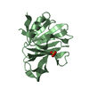
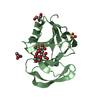
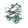
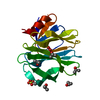
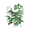
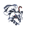
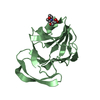
#1: Journal: Acta Crystallogr.,Sect.F / Year: 2005 Title: Cloning, expression, purification, crystallization and preliminary X-ray diffraction analysis of the VP8* carbohydrate-binding protein of the human rotavirus strain Wa Authors: Kraschnefski, M.J. / Scott, S.A. / Holloway, G. / Coulson, B.S. / von Itzstein, M. / Blanchard, H.
History
Deposition
Aug 16, 2006
Deposition site: PDBJ / Processing site: PDBJ
Revision 1.0
Apr 3, 2007
Provider: repository / Type: Initial release
Revision 1.1
Apr 1, 2008
Group: Version format compliance
Revision 1.2
Jul 13, 2011
Group: Non-polymer description / Version format compliance
Type: MAR CCD 165 mm / Detector: CCD / Date: Jul 16, 2004 / Details: mirrors
Radiation
Monochromator: silicon 111 / Protocol: SINGLE WAVELENGTH / Monochromatic (M) / Laue (L): M / Scattering type: x-ray
Radiation wavelength
Wavelength: 0.9794 Å / Relative weight: 1
Reflection
Resolution: 2.5→64.6 Å / Num. obs: 8135
-
Processing
Software
Name
Version
Classification
REFMAC
5.2.0019
refinement
XNEMO
datacollection
FIP
BM30A
datacollection
MOSFLM
datareduction
CCP4
(SCALA)
datascaling
AMoRE
phasing
Refinement
Method to determine structure: MOLECULAR REPLACEMENT Starting model: Homology model based on the structure of porcine CRW-8 VP8* that we have solved. Resolution: 2.5→64.55 Å / Cor.coef. Fo:Fc: 0.952 / Cor.coef. Fo:Fc free: 0.91 / SU B: 7.301 / SU ML: 0.163 / Cross valid method: THROUGHOUT / σ(F): 2 / ESU R: 0.402 / ESU R Free: 0.263 / Stereochemistry target values: MAXIMUM LIKELIHOOD / Details: HYDROGENS HAVE BEEN ADDED IN THE RIDING POSITIONS
Rfactor
Num. reflection
% reflection
Selection details
Rfree
0.23508
355
4.5 %
RANDOM
Rwork
0.17718
-
-
-
obs
0.17973
7491
96.54 %
-
all
-
8114
-
-
Solvent computation
Ion probe radii: 0.8 Å / Shrinkage radii: 0.8 Å / VDW probe radii: 1.4 Å / Solvent model: MASK
Displacement parameters
Biso mean: 31.888 Å2
Baniso -1
Baniso -2
Baniso -3
1-
1.36 Å2
0.68 Å2
0 Å2
2-
-
1.36 Å2
0 Å2
3-
-
-
-2.04 Å2
Refinement step
Cycle: LAST / Resolution: 2.5→64.55 Å
Protein
Nucleic acid
Ligand
Solvent
Total
Num. atoms
1300
0
20
93
1413
Refine LS restraints
Refine-ID
Type
Dev ideal
Dev ideal target
Number
X-RAY DIFFRACTION
r_bond_refined_d
0.008
0.022
1356
X-RAY DIFFRACTION
r_angle_refined_deg
1.106
1.925
1846
X-RAY DIFFRACTION
r_dihedral_angle_1_deg
6.653
5
161
X-RAY DIFFRACTION
r_dihedral_angle_2_deg
33.678
24.366
71
X-RAY DIFFRACTION
r_dihedral_angle_3_deg
16.865
15
209
X-RAY DIFFRACTION
r_dihedral_angle_4_deg
12.765
15
8
X-RAY DIFFRACTION
r_chiral_restr
0.082
0.2
200
X-RAY DIFFRACTION
r_gen_planes_refined
0.003
0.02
1057
X-RAY DIFFRACTION
r_nbd_refined
0.197
0.2
583
X-RAY DIFFRACTION
r_nbtor_refined
0.307
0.2
909
X-RAY DIFFRACTION
r_xyhbond_nbd_refined
0.115
0.2
102
X-RAY DIFFRACTION
r_symmetry_vdw_refined
0.206
0.2
40
X-RAY DIFFRACTION
r_symmetry_hbond_refined
0.169
0.2
10
X-RAY DIFFRACTION
r_mcbond_it
0.528
1.5
829
X-RAY DIFFRACTION
r_mcangle_it
0.949
2
1322
X-RAY DIFFRACTION
r_scbond_it
1.193
3
608
X-RAY DIFFRACTION
r_scangle_it
1.973
4.5
523
LS refinement shell
Resolution: 2.5→2.565 Å / Total num. of bins used: 20
Rfactor
Num. reflection
% reflection
Rfree
0.237
23
-
Rwork
0.271
533
-
obs
-
-
97.2 %
+
About Yorodumi
-
News
-
Feb 9, 2022. New format data for meta-information of EMDB entries
New format data for meta-information of EMDB entries
Version 3 of the EMDB header file is now the official format.
The previous official version 1.9 will be removed from the archive.
In the structure databanks used in Yorodumi, some data are registered as the other names, "COVID-19 virus" and "2019-nCoV". Here are the details of the virus and the list of structure data.
Jan 31, 2019. EMDB accession codes are about to change! (news from PDBe EMDB page)
EMDB accession codes are about to change! (news from PDBe EMDB page)
The allocation of 4 digits for EMDB accession codes will soon come to an end. Whilst these codes will remain in use, new EMDB accession codes will include an additional digit and will expand incrementally as the available range of codes is exhausted. The current 4-digit format prefixed with “EMD-” (i.e. EMD-XXXX) will advance to a 5-digit format (i.e. EMD-XXXXX), and so on. It is currently estimated that the 4-digit codes will be depleted around Spring 2019, at which point the 5-digit format will come into force.
The EM Navigator/Yorodumi systems omit the EMD- prefix.
Related info.:Q: What is EMD? / ID/Accession-code notation in Yorodumi/EM Navigator
Yorodumi is a browser for structure data from EMDB, PDB, SASBDB, etc.
This page is also the successor to EM Navigator detail page, and also detail information page/front-end page for Omokage search.
The word "yorodu" (or yorozu) is an old Japanese word meaning "ten thousand". "mi" (miru) is to see.
Related info.:EMDB / PDB / SASBDB / Comparison of 3 databanks / Yorodumi Search / Aug 31, 2016. New EM Navigator & Yorodumi / Yorodumi Papers / Jmol/JSmol / Function and homology information / Changes in new EM Navigator and Yorodumi
 Movie
Movie Controller

 Yorodumi
 Open data
 Basic information
 Components
 Keywords
 Function and homology information
 Human rotavirus A
 X-RAY DIFFRACTION /
  Authors
 Citation
 Structure visualization
 Downloads & links
 Other downloads
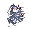


 PDBj
PDBj
 Assembly
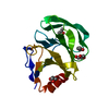


 Mass: 92.094 Da / Num. of mol.: 2 / Source method: obtained synthetically / Formula: C3H8O3
 Mass: 60.095 Da / Num. of mol.: 2 / Source method: obtained synthetically / Formula: C3H8O
 Mass: 18.015 Da / Num. of mol.: 93 / Source method: isolated from a natural source / Formula: H2O
 Sample preparation
 / Beamline: BM30A / Wavelength: 0.9794 Å
 Processing