[English] 日本語
Yorodumi
- PDB-2i2s: Crystal Structure of the porcine CRW-8 rotavirus VP8* carbohydrat... -

+
Open data


ID or keywords:

Loading...

-
Basic information

Entry
Database: PDB / ID: 2i2s
TitleCrystal Structure of the porcine CRW-8 rotavirus VP8* carbohydrate-recognising domain
ComponentsOuter capsid protein VP4
KeywordsVIRAL PROTEIN / beta-sandwich
Function / homology
Function and homology information


host cell rough endoplasmic reticulum / host cytoskeleton / viral outer capsid / permeabilization of host organelle membrane involved in viral entry into host cell / symbiont entry into host cell via permeabilization of inner membrane / host cell endoplasmic reticulum-Golgi intermediate compartment / virion attachment to host cell / host cell plasma membrane / membrane
Similarity search - Function
Haemagglutinin outer capsid protein VP4, concanavalin-like domain / Outer Capsid protein VP4 (Hemagglutinin) Concanavalin-like domain / Rotavirus VP4 helical domain / Rotavirus VP4 helical domain / Outer capsid protein VP4 / Rotavirus VP4, membrane interaction domain superfamily / Rotavirus VP4, membrane interaction domain / Rotavirus VP4 membrane interaction domain / Jelly Rolls - #200 / Concanavalin A-like lectin/glucanase domain superfamily ...Haemagglutinin outer capsid protein VP4, concanavalin-like domain / Outer Capsid protein VP4 (Hemagglutinin) Concanavalin-like domain / Rotavirus VP4 helical domain / Rotavirus VP4 helical domain / Outer capsid protein VP4 / Rotavirus VP4, membrane interaction domain superfamily / Rotavirus VP4, membrane interaction domain / Rotavirus VP4 membrane interaction domain / Jelly Rolls - #200 / Concanavalin A-like lectin/glucanase domain superfamily / Jelly Rolls / Sandwich / Mainly Beta
Similarity search - Domain/homology
2-O-methyl-5-N-acetyl-alpha-D-neuraminic acid / Outer capsid protein VP4 / Outer capsid protein VP4
Similarity search - Component
Biological speciesPorcine rotavirus
MethodX-RAY DIFFRACTION / SYNCHROTRON / MOLECULAR REPLACEMENT / Resolution: 2.3 Å
AuthorsBlanchard, H.
Citation
Journal: J.Mol.Biol. / Year: 2007
Title: Insight into Host Cell Carbohydrate-recognition by Human and Porcine Rotavirus from Crystal Structures of the Virion Spike Associated Carbohydrate-binding Domain (VP8*)
Authors: Blanchard, H. / Yu, X. / Coulson, B.S. / von Itzstein, M.
#1: Journal: Acta Crystallogr.,Sect.F / Year: 2005
Title: Crystallization and preliminary X-ray diffraction analysis of the sialic acid-binding domain (VP8*) of porcine rotavirus strain CRW-8
Authors: Scott, S.A. / Holloway, G. / Coulson, B.S. / Szyczew, A.J. / Kiefel, M.J. / von Itzstein, M. / Blanchard, H.
History
DepositionAug 16, 2006Deposition site: RCSB / Processing site: PDBJ
Revision 1.0Apr 3, 2007Provider: repository / Type: Initial release
Revision 1.1May 1, 2008Group: Version format compliance
Revision 1.2Jul 13, 2011Group: Non-polymer description / Version format compliance
Revision 1.3Jul 29, 2020Group: Data collection / Derived calculations / Structure summary
Category: chem_comp / entity ...chem_comp / entity / pdbx_chem_comp_identifier / pdbx_entity_nonpoly / pdbx_struct_conn_angle / struct_conn / struct_site / struct_site_gen
Item: _chem_comp.mon_nstd_flag / _chem_comp.name ..._chem_comp.mon_nstd_flag / _chem_comp.name / _chem_comp.type / _entity.pdbx_description / _pdbx_entity_nonpoly.name / _pdbx_struct_conn_angle.ptnr1_auth_comp_id / _pdbx_struct_conn_angle.ptnr1_auth_seq_id / _pdbx_struct_conn_angle.ptnr1_label_asym_id / _pdbx_struct_conn_angle.ptnr1_label_atom_id / _pdbx_struct_conn_angle.ptnr1_label_comp_id / _pdbx_struct_conn_angle.ptnr1_label_seq_id / _pdbx_struct_conn_angle.ptnr3_auth_comp_id / _pdbx_struct_conn_angle.ptnr3_auth_seq_id / _pdbx_struct_conn_angle.ptnr3_label_asym_id / _pdbx_struct_conn_angle.ptnr3_label_atom_id / _pdbx_struct_conn_angle.ptnr3_label_comp_id / _pdbx_struct_conn_angle.ptnr3_label_seq_id / _pdbx_struct_conn_angle.value / _struct_conn.pdbx_dist_value / _struct_conn.ptnr1_auth_asym_id / _struct_conn.ptnr1_auth_comp_id / _struct_conn.ptnr1_auth_seq_id / _struct_conn.ptnr1_label_asym_id / _struct_conn.ptnr1_label_atom_id / _struct_conn.ptnr1_label_comp_id / _struct_conn.ptnr1_label_seq_id / _struct_conn.ptnr2_auth_asym_id / _struct_conn.ptnr2_auth_comp_id / _struct_conn.ptnr2_auth_seq_id / _struct_conn.ptnr2_label_asym_id / _struct_conn.ptnr2_label_atom_id / _struct_conn.ptnr2_label_comp_id / _struct_conn.ptnr2_label_seq_id
Description: Carbohydrate remediation / Provider: repository / Type: Remediation
Revision 1.4Mar 13, 2024Group: Data collection / Database references / Structure summary
Category: chem_comp / chem_comp_atom ...chem_comp / chem_comp_atom / chem_comp_bond / database_2
Item: _chem_comp.pdbx_synonyms / _database_2.pdbx_DOI / _database_2.pdbx_database_accession
Revision 1.5Apr 3, 2024Group: Refinement description / Category: pdbx_initial_refinement_model

-
Structure visualization

Structure viewerMolecule:
MolmilJmol/JSmol

Downloads & links

-
Assembly

Deposited unit
A: Outer capsid protein VP4
B: Outer capsid protein VP4
hetero molecules


Theoretical massNumber of molelcules
Total (without water)38,52713
Polymers36,8992
Non-polymers1,62911
Water6,539363
1
A: Outer capsid protein VP4
hetero molecules


Theoretical massNumber of molelcules
Total (without water)19,3336
Polymers18,4491
Non-polymers8845
Water181
TypeNameSymmetry operationNumber
identity operation1_555x,y,z1
2
B: Outer capsid protein VP4
hetero molecules


Theoretical massNumber of molelcules
Total (without water)19,1947
Polymers18,4491
Non-polymers7456
Water181
TypeNameSymmetry operationNumber
identity operation1_555x,y,z1
Unit cell
Length a, b, c (Å)53.929, 64.663, 109.886
Angle α, β, γ (deg.)90.00, 90.00, 90.00
Int Tables number19
Space group name H-MP212121
Details
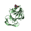
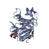
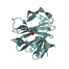
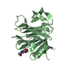
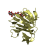
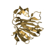
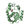
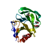
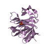
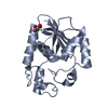
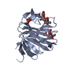
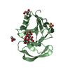
The biological unit is a monomer of VP8* which forms part of the rotavirus spike protein (VP4). The spike protein is considered to be formed of a dimer (though some evidence suggests a trimer) of VP4. The crystallographic asymmetric unit contains two molecules of VP8*.

-
Components

-
Protein / Sugars , 2 types, 5 molecules AB

#1: Protein Outer capsid protein VP4 / spike protein


Mass: 18449.479 Da / Num. of mol.: 2 / Fragment: VP8* domain
Source method: isolated from a genetically manipulated source
Source: (gene. exp.) Porcine rotavirus / Species: Rotavirus C / Strain: strain CRW-8 / Plasmid: pGex-VP8*(64-224) / Species (production host): Escherichia coli / Production host: Escherichia coli BL21(DE3) (bacteria) / Strain (production host): Bl21 DE3 / References: UniProt: P11114, UniProt: P0C6Y8*PLUS
#4: Sugar ChemComp-MNA / 2-O-methyl-5-N-acetyl-alpha-D-neuraminic acid / 2-O-METHYL-5-N-ACETYL-ALPHA-D- NEURAMINIC ACID


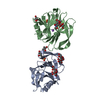
Type: D-saccharide / Mass: 323.296 Da / Num. of mol.: 3 / Source method: obtained synthetically / Formula: C12H21NO9
IdentifierTypeProgram
2-O-methyl-5-N-acetyl-a-D-neuraminic acidIUPAC CARBOHYDRATE SYMBOLPDB-CARE 1.0

-
Non-polymers , 5 types, 371 molecules

#2: Chemical ChemComp-SO4 / SULFATE ION / Sulfate


Mass: 96.063 Da / Num. of mol.: 2 / Source method: obtained synthetically / Formula: SO4
#3: Chemical ChemComp-NA / SODIUM ION


Mass: 22.990 Da / Num. of mol.: 2 / Source method: obtained synthetically / Formula: Na
#5: Chemical ChemComp-MPD / (4S)-2-METHYL-2,4-PENTANEDIOL / 2-Methyl-2,4-pentanediol


Mass: 118.174 Da / Num. of mol.: 2 / Source method: obtained synthetically / Formula: C6H14O2 / Comment: precipitant*YM
#6: Chemical ChemComp-GOL / GLYCEROL / GLYCERIN / PROPANE-1,2,3-TRIOL / Glycerol


Mass: 92.094 Da / Num. of mol.: 2 / Source method: obtained synthetically / Formula: C3H8O3
#7: Water ChemComp-HOH / water / Water


Mass: 18.015 Da / Num. of mol.: 363 / Source method: isolated from a natural source / Formula: H2O

-
Experimental details

-
Experiment

ExperimentMethod: X-RAY DIFFRACTION / Number of used crystals: 1

-
Sample preparation

CrystalDensity Matthews: 2.59 Å3/Da / Density % sol: 52.6 %
Crystal growTemperature: 295 K / Method: vapor diffusion, hanging drop / pH: 7.5
Details: 70% MPD, 0.1M HEPES pH 7.5, methyl-alpha-D-N-acetylneuraminide , VAPOR DIFFUSION, HANGING DROP, temperature 295K

-
Data collection

DiffractionMean temperature: 100 K
Diffraction sourceSource: SYNCHROTRON / Site: ESRF / Beamline: BM30A / Wavelength: 0.9794 Å
DetectorType: MAR CCD 165 mm / Detector: CCD / Date: Jul 16, 2004 / Details: mirrors
RadiationMonochromator: silicon 111 / Protocol: SINGLE WAVELENGTH / Monochromatic (M) / Laue (L): M / Scattering type: x-ray
Radiation wavelengthWavelength: 0.9794 Å / Relative weight: 1
ReflectionResolution: 2.3→55.9 Å / Num. obs: 17704

-
Processing

Software
NameVersionClassification
REFMAC5.2.0019refinement
XNEWMOdata collection
FIPBM30Adata collection
MOSFLMdata reduction
CCP4(SCALA)data scaling
AMoREphasing
RefinementMethod to determine structure: MOLECULAR REPLACEMENT
Starting model: Homology model based on the structure of Rhesus rotavirus VP8*

Resolution: 2.3→20 Å / Cor.coef. Fo:Fc: 0.955 / Cor.coef. Fo:Fc free: 0.913 / SU B: 5.307 / SU ML: 0.133 / Cross valid method: THROUGHOUT / σ(F): 0 / ESU R: 0.32 / ESU R Free: 0.221 / Stereochemistry target values: MAXIMUM LIKELIHOOD / Details: HYDROGENS HAVE BEEN ADDED IN THE RIDING POSITIONS
RfactorNum. reflection% reflectionSelection details
Rfree0.21952 901 5.1 %RANDOM
Rwork0.16256 ---
all0.183 17658 --
obs0.1654 16756 99.77 %-
Solvent computationIon probe radii: 0.8 Å / Shrinkage radii: 0.8 Å / VDW probe radii: 1.4 Å / Solvent model: MASK
Displacement parametersBiso mean: 21.243 Å2
Baniso -1Baniso -2Baniso -3
1--1 Å20 Å20 Å2
2--1.74 Å20 Å2
3----0.74 Å2
Refinement stepCycle: LAST / Resolution: 2.3→20 Å
ProteinNucleic acidLigandSolventTotal
Num. atoms2606 0 106 363 3075
Refine LS restraints
Refine-IDTypeDev idealDev ideal targetNumber
X-RAY DIFFRACTIONr_bond_refined_d0.0070.0222796
X-RAY DIFFRACTIONr_angle_refined_deg1.061.9743842
X-RAY DIFFRACTIONr_dihedral_angle_1_deg6.1085324
X-RAY DIFFRACTIONr_dihedral_angle_2_deg34.63724.88125
X-RAY DIFFRACTIONr_dihedral_angle_3_deg12.93415413
X-RAY DIFFRACTIONr_dihedral_angle_4_deg10.175158
X-RAY DIFFRACTIONr_chiral_restr0.0630.2447
X-RAY DIFFRACTIONr_gen_planes_refined0.0030.022098
X-RAY DIFFRACTIONr_nbd_refined0.1870.21241
X-RAY DIFFRACTIONr_nbtor_refined0.3060.21926
X-RAY DIFFRACTIONr_xyhbond_nbd_refined0.1230.2353
X-RAY DIFFRACTIONr_metal_ion_refined0.080.26
X-RAY DIFFRACTIONr_symmetry_vdw_refined0.1380.259
X-RAY DIFFRACTIONr_symmetry_hbond_refined0.1590.236
X-RAY DIFFRACTIONr_mcbond_it0.3061.51633
X-RAY DIFFRACTIONr_mcangle_it0.59322704
X-RAY DIFFRACTIONr_scbond_it0.89231217
X-RAY DIFFRACTIONr_scangle_it1.434.51138
LS refinement shellResolution: 2.3→2.36 Å / Total num. of bins used: 20
RfactorNum. reflection% reflection
Rfree0.223 72 -
Rwork0.178 1212 -
obs--99.92 %

+
About Yorodumi

-
News

-
Feb 9, 2022. New format data for meta-information of EMDB entries

New format data for meta-information of EMDB entries

  • Version 3 of the EMDB header file is now the official format.
  • The previous official version 1.9 will be removed from the archive.

Related info.:EMDB header

External links:wwPDB to switch to version 3 of the EMDB data model

-
Aug 12, 2020. Covid-19 info

Covid-19 info

URL: https://pdbj.org/emnavi/covid19.php

New page: Covid-19 featured information page in EM Navigator.

Related info.:Covid-19 info / Mar 5, 2020. Novel coronavirus structure data

+
Mar 5, 2020. Novel coronavirus structure data

Novel coronavirus structure data

Related info.:Yorodumi Speices / Aug 12, 2020. Covid-19 info

External links:COVID-19 featured content - PDBj / Molecule of the Month (242):Coronavirus Proteases

+
Jan 31, 2019. EMDB accession codes are about to change! (news from PDBe EMDB page)

EMDB accession codes are about to change! (news from PDBe EMDB page)

  • The allocation of 4 digits for EMDB accession codes will soon come to an end. Whilst these codes will remain in use, new EMDB accession codes will include an additional digit and will expand incrementally as the available range of codes is exhausted. The current 4-digit format prefixed with “EMD-” (i.e. EMD-XXXX) will advance to a 5-digit format (i.e. EMD-XXXXX), and so on. It is currently estimated that the 4-digit codes will be depleted around Spring 2019, at which point the 5-digit format will come into force.
  • The EM Navigator/Yorodumi systems omit the EMD- prefix.

Related info.:Q: What is EMD? / ID/Accession-code notation in Yorodumi/EM Navigator

External links:EMDB Accession Codes are Changing Soon! / Contact to PDBj

+
Jul 12, 2017. Major update of PDB

Major update of PDB

  • wwPDB released updated PDB data conforming to the new PDBx/mmCIF dictionary.
  • This is a major update changing the version number from 4 to 5, and with Remediation, in which all the entries are updated.
  • In this update, many items about electron microscopy experimental information are reorganized (e.g. em_software).
  • Now, EM Navigator and Yorodumi are based on the updated data.

External links:wwPDB Remediation / Enriched Model Files Conforming to OneDep Data Standards Now Available in the PDB FTP Archive

-
Yorodumi

Thousand views of thousand structures

  • Yorodumi is a browser for structure data from EMDB, PDB, SASBDB, etc.
  • This page is also the successor to EM Navigator detail page, and also detail information page/front-end page for Omokage search.
  • The word "yorodu" (or yorozu) is an old Japanese word meaning "ten thousand". "mi" (miru) is to see.

Related info.:EMDB / PDB / SASBDB / Comparison of 3 databanks / Yorodumi Search / Aug 31, 2016. New EM Navigator & Yorodumi / Yorodumi Papers / Jmol/JSmol / Function and homology information / Changes in new EM Navigator and Yorodumi

Read more