 Movie
Movie Controller


+ Open data

- Basic information
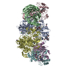
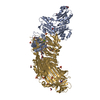
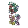
| Entry | Database: PDB / ID: 6pdt | ||||||
|---|---|---|---|---|---|---|---|
| Title | cryoEM structure of yeast glucokinase filament | ||||||
 Components Components | Glucokinase-1 | ||||||
 Keywords Keywords | TRANSFERASE / filament | ||||||
| Function / homology |  Function and homology information Function and homology informationSynthesis of GDP-mannose / glucokinase / Glycolysis / fructokinase activity / Regulation of Glucokinase by Glucokinase Regulatory Protein / glucokinase activity / mannose metabolic process / glucose 6-phosphate metabolic process / D-glucose binding / Neutrophil degranulation ...Synthesis of GDP-mannose / glucokinase / Glycolysis / fructokinase activity / Regulation of Glucokinase by Glucokinase Regulatory Protein / glucokinase activity / mannose metabolic process / glucose 6-phosphate metabolic process / D-glucose binding / Neutrophil degranulation / intracellular glucose homeostasis / glycolytic process / glucose metabolic process / mitochondrion / ATP binding / plasma membrane / cytosol Similarity search - Function | ||||||
| Biological species |  | ||||||
| Method | ELECTRON MICROSCOPY / helical reconstruction / cryo EM / Resolution: 3.8 Å | ||||||
| Model details | MODEL GENERATED BY ROSETTA VERSION 2019.21.post.dev+31.master.d8f9b4a90a8 | ||||||
 Authors Authors | Lynch, E.M. / Dosey, A.M. / Farrell, D.P. / Stoddard, P.R. / Kollman, J.M. | ||||||
 Citation Citation | Journal: Science / Year: 2020 Title: Polymerization in the actin ATPase clan regulates hexokinase activity in yeast. Authors: Patrick R Stoddard / Eric M Lynch / Daniel P Farrell / Annie M Dosey / Frank DiMaio / Tom A Williams / Justin M Kollman / Andrew W Murray / Ethan C Garner /   Abstract: The actin fold is found in cytoskeletal polymers, chaperones, and various metabolic enzymes. Many actin-fold proteins, such as the carbohydrate kinases, do not polymerize. We found that Glk1, a ...The actin fold is found in cytoskeletal polymers, chaperones, and various metabolic enzymes. Many actin-fold proteins, such as the carbohydrate kinases, do not polymerize. We found that Glk1, a glucokinase, forms two-stranded filaments with ultrastructure that is distinct from that of cytoskeletal polymers. In cells, Glk1 polymerized upon sugar addition and depolymerized upon sugar withdrawal. Polymerization inhibits enzymatic activity; the Glk1 monomer-polymer equilibrium sets a maximum rate of glucose phosphorylation regardless of Glk1 concentration. A mutation that eliminated Glk1 polymerization alleviated concentration-dependent enzyme inhibition. Yeast containing nonpolymerizing Glk1 were less fit when growing on sugars and more likely to die when refed glucose. Glk1 polymerization arose independently from other actin-related filaments and may allow yeast to rapidly modulate glucokinase activity as nutrient availability changes. | ||||||
| History |
|
- Structure visualization
| Movie |
Movie viewer |
|---|---|
| Structure viewer | Molecule: MolmilJmol/JSmol |
- Downloads & links
-Download
| PDBx/mmCIF format | 6pdt.cif.gz | 636 KB | Display | PDBx/mmCIF format |
|---|---|---|---|---|
| PDB format | pdb6pdt.ent.gz | 524.9 KB | Display | PDB format |
| PDBx/mmJSON format | 6pdt.json.gz | Tree view | PDBx/mmJSON format | |
| Others |  Other downloads Other downloads |
-Validation report
| Arichive directory | https://data.pdbj.org/pub/pdb/validation_reports/pd/6pdtftp://data.pdbj.org/pub/pdb/validation_reports/pd/6pdt | HTTPS FTP |
|---|
-Related structure data
| Related structure data | 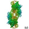 20309MC  6p4xC M: map data used to model this data C: citing same article ( |
|---|---|
| Similar structure data |


-Links
 PDBj
PDBj


- Assembly
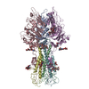
| Deposited unit | 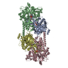
|
|---|---|
| 1 |
|
-Components
| #1: Protein | Mass: 55446.258 Da / Num. of mol.: 4 Source method: isolated from a genetically manipulated source Source: (gene. exp.) Strain: ATCC 204508 / S288c / Gene: GLK1, HOR3, YCL040W, YCL312, YCL40W / Production host:  #2: Sugar | ChemComp-GLC /   Type: D-saccharide, alpha linking / Mass: 180.156 Da / Num. of mol.: 4 Type: D-saccharide, alpha linking / Mass: 180.156 Da / Num. of mol.: 4Source method: isolated from a genetically manipulated source Formula: C6H12O6 #3: Chemical | ChemComp-MG /   Mass: 24.305 Da / Num. of mol.: 4 / Source method: obtained synthetically / Formula: Mg Mass: 24.305 Da / Num. of mol.: 4 / Source method: obtained synthetically / Formula: Mg#4: Chemical | ChemComp-ATP /   Mass: 507.181 Da / Num. of mol.: 4 / Source method: obtained synthetically / Formula: C10H16N5O13P3 / Comment: ATP, energy-carrying molecule*YM Mass: 507.181 Da / Num. of mol.: 4 / Source method: obtained synthetically / Formula: C10H16N5O13P3 / Comment: ATP, energy-carrying molecule*YMHas ligand of interest | N | Has protein modification | Y | |
|---|
-Experimental details
-Experiment
| Experiment | Method: ELECTRON MICROSCOPY |
|---|---|
| EM experiment | Aggregation state: FILAMENT / 3D reconstruction method: helical reconstruction |
- Sample preparation
| Component | Name: glucokinase-1 / Type: COMPLEX / Entity ID: #1 / Source: RECOMBINANT |
|---|---|
| Molecular weight | Experimental value: NO |
| Source (natural) | Organism: |
| Source (recombinant) | Organism: |
| Buffer solution | pH: 7.5 |
| Specimen | Embedding applied: NO / Shadowing applied: NO / Staining applied: NO / Vitrification applied: YES |
| Vitrification | Cryogen name: ETHANE / Humidity: 100 % |
- Electron microscopy imaging
| Experimental equipment |  Model: Titan Krios / Image courtesy: FEI Company |
|---|---|
| Microscopy | Model: FEI TITAN KRIOS |
| Electron gun | Electron source:  FIELD EMISSION GUN / Accelerating voltage: 300 kV / Illumination mode: FLOOD BEAM FIELD EMISSION GUN / Accelerating voltage: 300 kV / Illumination mode: FLOOD BEAM |
| Electron lens | Mode: BRIGHT FIELD |
| Image recording | Electron dose: 90 e/Å2 / Detector mode: SUPER-RESOLUTION / Film or detector model: GATAN K2 SUMMIT (4k x 4k) |
- Processing
| EM software |
| |||||||||||||||||||||||||||
|---|---|---|---|---|---|---|---|---|---|---|---|---|---|---|---|---|---|---|---|---|---|---|---|---|---|---|---|---|
| CTF correction | Type: PHASE FLIPPING AND AMPLITUDE CORRECTION | |||||||||||||||||||||||||||
| Helical symmerty | Angular rotation/subunit: 120.4 ° / Axial rise/subunit: 60.1 Å / Axial symmetry: D1 | |||||||||||||||||||||||||||
| 3D reconstruction | Resolution: 3.8 Å / Resolution method: FSC 0.143 CUT-OFF / Num. of particles: 56778 / Symmetry type: HELICAL |
