 Movie
Movie Controller

[English] 日本語
 Yorodumi
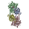
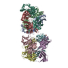
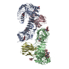
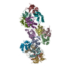
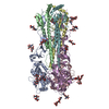
- PDB-1flc: X-RAY STRUCTURE OF THE HAEMAGGLUTININ-ESTERASE-FUSION GLYCOPROTEI... -
+ Open data

- Basic information
| Entry | Database: PDB / ID: 1flc | |||||||||
|---|---|---|---|---|---|---|---|---|---|---|
| Title | X-RAY STRUCTURE OF THE HAEMAGGLUTININ-ESTERASE-FUSION GLYCOPROTEIN OF INFLUENZA C VIRUS | |||||||||
 Components Components | (HAEMAGGLUTININ-ESTERASE-FUSION GLYCOPROTEIN) x 2 | |||||||||
 Keywords Keywords | HYDROLASE / ESTERASE / RECEPTOR BINDING / MEMBRANE FUSION / VIRUS / INFLUENZA | |||||||||
| Function / homology |  Function and homology information Function and homology informationsialate 9-O-acetylesterase activity / sialate 4-O-acetylesterase activity / sialate O-acetylesterase / viral budding from plasma membrane / host cell surface receptor binding / endocytosis involved in viral entry into host cell / fusion of virus membrane with host plasma membrane / fusion of virus membrane with host endosome membrane / viral envelope / virion attachment to host cell ...sialate 9-O-acetylesterase activity / sialate 4-O-acetylesterase activity / sialate O-acetylesterase / viral budding from plasma membrane / host cell surface receptor binding / endocytosis involved in viral entry into host cell / fusion of virus membrane with host plasma membrane / fusion of virus membrane with host endosome membrane / viral envelope / virion attachment to host cell / host cell plasma membrane / virion membrane / membrane Similarity search - Function | |||||||||
| Biological species |  Influenza C virus Influenza C virus | |||||||||
| Method |  X-RAY DIFFRACTION / SYNCHROTRON / SIR / Resolution: 3.2 Å X-RAY DIFFRACTION / SYNCHROTRON / SIR / Resolution: 3.2 Å | |||||||||
 Authors Authors | Rosenthal, P.B. / Zhang, X. / Formanowski, F. / Fitz, W. / Wong, C.H. / Meier-Ewert, H. / Skehel, J.J. / Wiley, D.C. | |||||||||
 Citation Citation | Journal: Nature / Year: 1998 Title: Structure of the haemagglutinin-esterase-fusion glycoprotein of influenza C virus. Authors: Rosenthal, P.B. / Zhang, X. | |||||||||
| History |
|
- Structure visualization
| Structure viewer | Molecule: MolmilJmol/JSmol |
|---|
- Downloads & links
-Download
| PDBx/mmCIF format | 1flc.cif.gz | 354.2 KB | Display | PDBx/mmCIF format |
|---|---|---|---|---|
| PDB format | pdb1flc.ent.gz | 294 KB | Display | PDB format |
| PDBx/mmJSON format | 1flc.json.gz | Tree view | PDBx/mmJSON format | |
| Others |  Other downloads Other downloads |
-Validation report
| Arichive directory | https://data.pdbj.org/pub/pdb/validation_reports/fl/1flcftp://data.pdbj.org/pub/pdb/validation_reports/fl/1flc | HTTPS FTP |
|---|
-Related structure data
| Similar structure data |
|---|


-Links
 PDBj
PDBj


- Assembly
| Deposited unit | 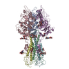
| ||||||||||||
|---|---|---|---|---|---|---|---|---|---|---|---|---|---|
| 1 |
| ||||||||||||
| Unit cell |
| ||||||||||||
| Noncrystallographic symmetry (NCS) | NCS oper:
|
-Components
| #1: Protein | Mass: 48230.699 Da / Num. of mol.: 3 / Fragment: HEF1 Source method: isolated from a genetically manipulated source Source: (gene. exp.) Influenza C virus (C/Johannesburg/1/66)Genus: Influenzavirus C / Species: Influenza C virus / Strain: C/JOHANNESBURG/1/66 / References: UniProt: P07975 #2: Protein | Mass: 18977.541 Da / Num. of mol.: 3 / Fragment: HEF2 Source method: isolated from a genetically manipulated source Source: (gene. exp.) Influenza C virus (C/Johannesburg/1/66)Genus: Influenzavirus C / Species: Influenza C virus / Strain: C/JOHANNESBURG/1/66 / References: GenBank: 325318, UniProt: P07975*PLUS #3: Polysaccharide | beta-D-mannopyranose-(1-4)-2-acetamido-2-deoxy-beta-D-glucopyranose-(1-4)-2-acetamido-2-deoxy-beta- ...beta-D-mannopyranose-(1-4)-2-acetamido-2-deoxy-beta-D-glucopyranose-(1-4)-2-acetamido-2-deoxy-beta-D-glucopyranose Source method: isolated from a genetically manipulated source #4: Polysaccharide | Source method: isolated from a genetically manipulated source #5: Polysaccharide | beta-D-mannopyranose-(1-4)-2-acetamido-2-deoxy-alpha-D-glucopyranose-(1-4)-2-acetamido-2-deoxy-beta- ...beta-D-mannopyranose-(1-4)-2-acetamido-2-deoxy-alpha-D-glucopyranose-(1-4)-2-acetamido-2-deoxy-beta-D-glucopyranose | Source method: isolated from a genetically manipulated source Has protein modification | Y | |
|---|
-Experimental details
-Experiment
| Experiment | Method: X-RAY DIFFRACTION |
|---|
- Sample preparation
| Crystal | Density Matthews: 6.2 Å3/Da / Density % sol: 76 % | ||||||||||||||||||||||||||||||||||||||||||||||||||||||||||||||||||
|---|---|---|---|---|---|---|---|---|---|---|---|---|---|---|---|---|---|---|---|---|---|---|---|---|---|---|---|---|---|---|---|---|---|---|---|---|---|---|---|---|---|---|---|---|---|---|---|---|---|---|---|---|---|---|---|---|---|---|---|---|---|---|---|---|---|---|---|
| Crystal grow | pH: 7.1 / Details: pH 7.10 | ||||||||||||||||||||||||||||||||||||||||||||||||||||||||||||||||||
| Crystal grow | *PLUS Method: vapor diffusion, hanging drop | ||||||||||||||||||||||||||||||||||||||||||||||||||||||||||||||||||
| Components of the solutions | *PLUS
|
-Data collection
| Diffraction | Mean temperature: 108 K |
|---|---|
| Diffraction source | Source: SYNCHROTRON / Site: CHESS  / Beamline: F1 / Wavelength: 0.91 / Beamline: F1 / Wavelength: 0.91 |
| Detector | Type: FUJI / Detector: IMAGE PLATE |
| Radiation | Protocol: SINGLE WAVELENGTH / Monochromatic (M) / Laue (L): M / Scattering type: x-ray |
| Radiation wavelength | Wavelength: 0.91 Å / Relative weight: 1 |
| Reflection | Resolution: 3.2→15 Å / Num. obs: 80900 / % possible obs: 99 % / Redundancy: 3.6 % / Biso Wilson estimate: 40 Å2 / Rsym value: 0.1 / Net I/σ(I): 12 |
| Reflection shell | Resolution: 3.2→10 Å / Redundancy: 3.6 % / Rmerge(I) obs: 0.1 / Mean I/σ(I) obs: 12 / Rsym value: 0.3 / % possible all: 91 |
| Reflection | *PLUS Rmerge(I) obs: 0.1 |
| Reflection shell | *PLUS % possible obs: 91 % |
- Processing
| Software |
| ||||||||||||||||||||||||||||||||||||||||||||||||||||||||||||
|---|---|---|---|---|---|---|---|---|---|---|---|---|---|---|---|---|---|---|---|---|---|---|---|---|---|---|---|---|---|---|---|---|---|---|---|---|---|---|---|---|---|---|---|---|---|---|---|---|---|---|---|---|---|---|---|---|---|---|---|---|---|
| Refinement | Method to determine structure: SIR / Resolution: 3.2→10 Å / Isotropic thermal model: RESTRAINED / Cross valid method: THROUGHOUT / σ(F): 2
| ||||||||||||||||||||||||||||||||||||||||||||||||||||||||||||
| Displacement parameters | Biso mean: 40 Å2 | ||||||||||||||||||||||||||||||||||||||||||||||||||||||||||||
| Refinement step | Cycle: LAST / Resolution: 3.2→10 Å
| ||||||||||||||||||||||||||||||||||||||||||||||||||||||||||||
| Refine LS restraints |
| ||||||||||||||||||||||||||||||||||||||||||||||||||||||||||||
| Refine LS restraints NCS | NCS model details: RESTRAINTS |
