 ムービー
ムービー コントローラー
 構造ビューア
 EMN検索について


-検索条件
-検索結果
検索 (著者・登録者: evans & g)の結果70件中、1から50件目までを表示しています

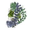
PDB-8p2w: 
Structure of human SIT1 (focussed map / refinement)

PDB-8p2z:
Structure of human SIT1 bound to L-pipecolate (focussed map / refinement)
PDB-8p30:
Structure of human SIT1:ACE2 complex (open PD conformation) bound to L-pipecolate

EMDB-17377:
Structure of human SIT1 (focussed map / refinement)

EMDB-17378:
Structure of human SIT1:ACE2 complex (open PD conformation)

EMDB-17379:
Structure of human SIT1:ACE2 complex (closed PD conformation)

EMDB-17380:
Structure of human SIT1 bound to L-pipecolate (focussed map / refinement)

EMDB-17381:
Structure of human SIT1:ACE2 complex (open PD conformation) bound to L-pipecolate
EMDB-17382:
Structure of human SIT1:ACE2 complex (closed PD conformation) bound to L-pipecolate

PDB-8p2x:
Structure of human SIT1:ACE2 complex (open PD conformation)

PDB-8p2y:
Structure of human SIT1:ACE2 complex (closed PD conformation)

PDB-8p31:
Structure of human SIT1:ACE2 complex (closed PD conformation) bound to L-pipecolate

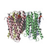
EMDB-28965:
Glutamine synthetase from Pseudomonas aeruginosa, filament double-unit in compressed conformation

PDB-8fbp:
Glutamine synthetase from Pseudomonas aeruginosa, filament double-unit in compressed conformation

EMDB-27738:
Negative stain EM map of the heterodimeric p110gamma-p84 complex

EMDB-14379:
Cryo-EM structure of an intact carboxysome - core layer
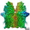
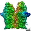
EMDB-14376:
Cryo-EM structure of the Cyanobium sp. PCC 7001 RuBisCO enzyme at 3.8 A resolution with C1 symmetry
PDB-8cmy:
Structure of the Cyanobium sp. PCC 7001 determined with C1 symmetry
EMDB-14377:
Cryo-EM structure of Cyanobium sp. PCC 7001 carboxysome shell
EMDB-14380:
Cryo-EM structure of the Cyanobium sp. PCC 7001 Carboxysome internal density outer layer
EMDB-14381:
Cryo-EM structure of the Cyanobium sp. PCC 7001 Carboxysome internal density middle layer
EMDB-14385:
Cryo-EM structure of an a-carboxysome RuBisCO enzyme at 2.9 A resolution

PDB-7yyo:
Cryo-EM structure of an a-carboxysome RuBisCO enzyme at 2.9 A resolution
EMDB-15905:
Cryo-EM structure of the E.coli 70S ribosome in complex with the antibiotic Myxovalargin B.
PDB-8b7y:
Cryo-EM structure of the E.coli 70S ribosome in complex with the antibiotic Myxovalargin B.

EMDB-15409:
C1 Cyanobacteria sp PCC 7001 RuBisCO

EMDB-14121:
Cryo-EM structure of the E.coli 50S ribosomal subunit in complex with the antibiotic Myxovalargin A.

PDB-7qq3:
Cryo-EM structure of the E.coli 50S ribosomal subunit in complex with the antibiotic Myxovalargin A.

EMDB-28551:
RMC-5552 in complex with mTORC1 and FKBP12

PDB-8era:
RMC-5552 in complex with mTORC1 and FKBP12

EMDB-28816:
Wild type P53 dimer structure from human cancer cells

EMDB-28817:
P53 monomer structure

PDB-8f2h:
Wild type P53 dimer structure from human cancer cells
PDB-8f2i:
P53 monomer structure
EMDB-14666:
Cryo-EM structure of Human ACE2 bound to a high-affinity SARS CoV-2 mutant

PDB-7zdq:
Cryo-EM structure of Human ACE2 bound to a high-affinity SARS CoV-2 mutant

EMDB-14517:
Chimera of AP2 with FCHO2 linker domain as a fusion on Cmu2 subunit

EMDB-14518:
Chimaera of AP2 with FCHO2 linker domain, N1-N2 enriched population

EMDB-14525:
AP2 adaptor protein recruited on the membrane in the presence of FCHO2 linker

EMDB-14526:
AP2 on the membrane without cargo peptide
PDB-7z5c:
Chimera of AP2 with FCHO2 linker domain as a fusion on Cmu2 subunit

EMDB-13035:
Cryo-EM structure of human TMEM45A
PDB-7oqz:
Cryo-EM structure of human TMEM45A
EMDB-11273:
Open-open state of the Bt1762-Bt1763 levan transport system

EMDB-11274:
Closed-closed state of the Bt1762-Bt1763 levan transport system
EMDB-11277:
Open-closed state of the Bt1762-Bt1763 levan transport system

PDB-6zlt:
Open-open state of the Bt1762-Bt1763 levan transport system

PDB-6zlu:
Closed-closed state of the Bt1762-Bt1763 levan transport system

PDB-6zm1:
Open-closed state of the Bt1762-Bt1763 levan transport system
EMDB-22827:
P53 tetramer from Glioblastoma
ページ:
 wwPDBはEMDBデータモデルのバージョン3へ移行します
