 Movie
Movie Controller


+ Open data

- Basic information
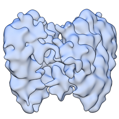
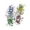
| Entry | Database: EMDB / ID: EMD-22827 | ||||||||||||
|---|---|---|---|---|---|---|---|---|---|---|---|---|---|
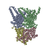
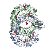
| Title | P53 tetramer from Glioblastoma | ||||||||||||
 Map data Map data | P53 tetramer from Glioblastoma | ||||||||||||
 Sample Sample |
| ||||||||||||
| Biological species |  Homo sapiens (human) Homo sapiens (human) | ||||||||||||
| Method | single particle reconstruction / cryo EM / Resolution: 7.0 Å | ||||||||||||
 Authors Authors | Solares MJ / Kelly DF | ||||||||||||
| Funding support |  United States, 3 items United States, 3 items
| ||||||||||||
 Citation Citation | Journal: Anal Chem / Year: 2020 Title: Microchip-Based Structure Determination of Disease-Relevant p53. Authors: Maria J Solares / G M Jonaid / William Y Luqiu / Yanping Liang / Madison C Evans / William J Dearnaley / Zhi Sheng / Deborah F Kelly / Abstract: The tumor suppressor protein TP53 (p53) plays a multifaceted role in all cells of the human body. Mutations in the gene are often involved in cancer induction and disease progression. Despite its ...The tumor suppressor protein TP53 (p53) plays a multifaceted role in all cells of the human body. Mutations in the gene are often involved in cancer induction and disease progression. Despite its important role in health and development, structural information for p53 remains incomplete. Here, we present a microchip-based technology to facilitate structural studies of p53 assemblies derived from human cancer cells. These devices do not introduce foreign sequences to the gene and maintain naturally occurring post-translational modifications. Using cryo-electron microscopy, structures for the p53 monomer (∼50 kDa) and tetramer (∼200 kDa) were resolved to ∼4.8 and ∼7 Å, respectively. These structures revealed new insights for flexible regions of p53 along with biologically relevant ubiquitination sites. Collectively, the convergence of nanotechnology tools and structural imaging builds a strong framework to understand the oncogenic impact of p53 in human tissues. | ||||||||||||
| History |
|
- Structure visualization
| Movie |
 Movie viewer Movie viewer |
|---|---|
| Structure viewer | EM map: SurfViewMolmilJmol/JSmol |
| Supplemental images |
- Downloads & links
-EMDB archive
| Map data | emd_22827.map.gz | 10.8 MB | EMDB map data format | |
|---|---|---|---|---|
| Header (meta data) | emd-22827-v30.xmlemd-22827.xml | 13.5 KB 13.5 KB | Display Display | EMDB header |
| Images |  emd_22827.png emd_22827.png | 178.6 KB | ||
| Archive directory |  http://ftp.pdbj.org/pub/emdb/structures/EMD-22827ftp://ftp.pdbj.org/pub/emdb/structures/EMD-22827 http://ftp.pdbj.org/pub/emdb/structures/EMD-22827ftp://ftp.pdbj.org/pub/emdb/structures/EMD-22827 | HTTPS FTP |
-Related structure data
| Similar structure data |
|---|

-Links
| EMDB pages | EMDB (EBI/PDBe) / EMDataResource |
|---|
-Map
| File | Download / File: emd_22827.map.gz / Format: CCP4 / Size: 30.5 MB / Type: IMAGE STORED AS FLOATING POINT NUMBER (4 BYTES) | ||||||||||||||||||||||||||||||||||||||||||||||||||||||||||||
|---|---|---|---|---|---|---|---|---|---|---|---|---|---|---|---|---|---|---|---|---|---|---|---|---|---|---|---|---|---|---|---|---|---|---|---|---|---|---|---|---|---|---|---|---|---|---|---|---|---|---|---|---|---|---|---|---|---|---|---|---|---|
| Annotation | P53 tetramer from Glioblastoma | ||||||||||||||||||||||||||||||||||||||||||||||||||||||||||||
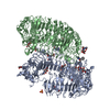
| Projections & slices | Image control
Images are generated by Spider. | ||||||||||||||||||||||||||||||||||||||||||||||||||||||||||||
| Voxel size | X=Y=Z: 1 Å | ||||||||||||||||||||||||||||||||||||||||||||||||||||||||||||
| Density |
| ||||||||||||||||||||||||||||||||||||||||||||||||||||||||||||
| Symmetry | Space group: 1 | ||||||||||||||||||||||||||||||||||||||||||||||||||||||||||||
| Details | EMDB XML:
CCP4 map header:
| ||||||||||||||||||||||||||||||||||||||||||||||||||||||||||||
 Z (Sec.)
 Y (Row.)
 X (Col.)


-Supplemental data
- Sample components
-Entire : Tetramer of p53 and DNA
| Entire | Name: Tetramer of p53 and DNA |
|---|---|
| Components |
|
-Supramolecule #1: Tetramer of p53 and DNA
| Supramolecule | Name: Tetramer of p53 and DNA / type: complex / ID: 1 / Parent: 0 / Macromolecule list: all / Details: isolated from glioblastoma cells |
|---|---|
| Source (natural) | Organism: Homo sapiens (human) / Organ: Brain / Tissue: epithelial / Location in cell: nucleus |
| Molecular weight | Theoretical: 200 KDa |
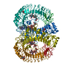
-Macromolecule #1: Tumor suppressor protein, P53
| Macromolecule | Name: Tumor suppressor protein, P53 / type: protein_or_peptide / ID: 1 / Enantiomer: DEXTRO |
|---|---|
| Source (natural) | Organism: Homo sapiens (human) / Organ: Brain / Tissue: epithelial |
| Sequence | String: MEEPQSDPSV EPPLSQETFS DLWKLLPENN VLSPLPSQAM DDLMLSPDDI EQWFTEDPGP DEAPRMPEA APPVAPAPAA PTPAAPAPAP SWPLSSSVPS QKTYQGSYGF RLGFLHSGTA K SVTCTYSP ALNKMFCQLA KTCPVQLWVD STPPPGTRVR AMAIYKQSQH ...String: MEEPQSDPSV EPPLSQETFS DLWKLLPENN VLSPLPSQAM DDLMLSPDDI EQWFTEDPGP DEAPRMPEA APPVAPAPAA PTPAAPAPAP SWPLSSSVPS QKTYQGSYGF RLGFLHSGTA K SVTCTYSP ALNKMFCQLA KTCPVQLWVD STPPPGTRVR AMAIYKQSQH MTEVVRRCPH HE RCSDSDG LAPPQHLIRV EGNLRVEYLD DRNTFRHSVV VPYEPPEVGS DCTTIHYNYM CNS SCMGGM NRRPILTIIT LEDSSGNLLG RNSFEVRVCA CPGRDRRTEE ENLRKKGEPH HELP PGSTK RALPNNTSSS PQPKKKPLDG EYFTLQIRGR ERFEMFRELN EALELKDAQA GKEPG GSRA HSSHLKSKKG QSTSRHKKLM FKTEGPDSD |
-Experimental details
-Structure determination
| Method | cryo EM |
|---|---|
 Processing Processing | single particle reconstruction |
| Aggregation state | particle |
-Sample preparation
| Concentration | 0.02 mg/mL |
|---|---|
| Buffer | pH: 7.2 Details: 20 mM HEPES, (pH 7.2), 140 mM NaCl, 2 mM CaCl2, 2 mM MgCl2, and 5 mM imidazole |
| Grid | Material: SILICON NITRIDE |
| Vitrification | Cryogen name: ETHANE / Chamber humidity: 90 % / Chamber temperature: 298 K / Instrument: FEI VITROBOT MARK III / Details: blotted for 6 seconds before plunging. |
| Details | Concentrated onto 25% Ni-NTA coating on silicon nitride microchips |
- Electron microscopy
| Microscope | TFS TALOS F200C |
|---|---|
| Image recording | Film or detector model: FEI CETA (4k x 4k) / Digitization - Sampling interval: 14.0 µm / Number grids imaged: 2 / Number real images: 150 / Average exposure time: 1.0 sec. / Average electron dose: 5.0 e/Å2 |
| Electron beam | Acceleration voltage: 200 kV / Electron source:  FIELD EMISSION GUN FIELD EMISSION GUN |
| Electron optics | C2 aperture diameter: 100.0 µm / Calibrated magnification: 92000 / Illumination mode: FLOOD BEAM / Imaging mode: BRIGHT FIELD / Cs: 2.0 mm / Nominal defocus max: 2.0 µm / Nominal defocus min: 0.5 µm / Nominal magnification: 92000 |
| Sample stage | Specimen holder model: GATAN 626 SINGLE TILT LIQUID NITROGEN CRYO TRANSFER HOLDER Cooling holder cryogen: NITROGEN |
| Experimental equipment |  Model: Tecnai F20 / Image courtesy: FEI Company |
+Image processing
-Atomic model buiding 1
| Initial model | PDB ID: |
|---|---|
| Refinement | Space: REAL / Protocol: RIGID BODY FIT |
