 Movie
Movie Controller

[English] 日本語
 Yorodumi
- PDB-8fbp: Glutamine synthetase from Pseudomonas aeruginosa, filament double... -
+ Open data

- Basic information
| Entry | Database: PDB / ID: 8fbp | ||||||
|---|---|---|---|---|---|---|---|
| Title | Glutamine synthetase from Pseudomonas aeruginosa, filament double-unit in compressed conformation | ||||||
 Components Components | Glutamine synthetase | ||||||
 Keywords Keywords | LIGASE / glutamine biosynthetic process nitrogen utilization / Structural Genomics / Seattle Structural Genomics Center for Infectious Disease / SSGCID | ||||||
| Function / homology |  Function and homology information Function and homology informationnitrogen utilization / glutamine synthetase / : / glutamine synthetase activity / ATP binding / membrane / metal ion binding / cytoplasm Similarity search - Function | ||||||
| Biological species |  Pseudomonas aeruginosa PAO1 (bacteria) Pseudomonas aeruginosa PAO1 (bacteria) | ||||||
| Method | ELECTRON MICROSCOPY / single particle reconstruction / cryo EM / Resolution: 2.8 Å | ||||||
 Authors Authors | Phan, I.Q. / Staker, B. / Shek, R. / Moser, T.H. / Evans, J.E. / van Voorhis, W.C. / Myler, P.J. / Seattle Structural Genomics Center for Infectious Disease (SSGCID) | ||||||
| Funding support |  United States, 1items United States, 1items
| ||||||
 Citation Citation | Journal: To Be Published Title: Glutamine synthetase from Pseudomonas aeruginosa, filament double-unit in compressed conformation Authors: Phan, I.Q. / Staker, B. / Shek, R. / Moser, T.H. / Evans, J.E. / van Voorhis, W.C. / Myler, P.J. | ||||||
| History |
|
- Structure visualization
| Structure viewer | Molecule: MolmilJmol/JSmol |
|---|
- Downloads & links
-Download
| PDBx/mmCIF format | 8fbp.cif.gz | 2.1 MB | Display | PDBx/mmCIF format |
|---|---|---|---|---|
| PDB format | pdb8fbp.ent.gz | Display | PDB format | |
| PDBx/mmJSON format | 8fbp.json.gz | Tree view | PDBx/mmJSON format | |
| Others |  Other downloads Other downloads |
-Validation report
| Arichive directory | https://data.pdbj.org/pub/pdb/validation_reports/fb/8fbpftp://data.pdbj.org/pub/pdb/validation_reports/fb/8fbp | HTTPS FTP |
|---|
-Related structure data
| Related structure data | 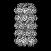 28965MC M: map data used to model this data C: citing same article ( |
|---|---|
| Similar structure data | |
| Other databases |
-Links
 PDBj
PDBj
- Assembly
| Deposited unit | 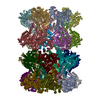
|
|---|---|
| 1 |
|
-Components
| #1: Protein | Mass: 53039.711 Da / Num. of mol.: 28 Source method: isolated from a genetically manipulated source Source: (gene. exp.) Pseudomonas aeruginosa PAO1 (bacteria) / Gene: glnA, PA5119Production host: References: UniProt: Q9HU65, glutamine synthetase |
|---|
-Experimental details
-Experiment
| Experiment | Method: ELECTRON MICROSCOPY |
|---|---|
| EM experiment | Aggregation state: FILAMENT / 3D reconstruction method: single particle reconstruction |
- Sample preparation
| Component | Name: P. aeruginosa GS filament. / Type: COMPLEX / Details: Filament formation during sample preparation. / Entity ID: all / Source: RECOMBINANT | |||||||||||||||||||||||||
|---|---|---|---|---|---|---|---|---|---|---|---|---|---|---|---|---|---|---|---|---|---|---|---|---|---|---|
| Source (natural) | Organism:  Pseudomonas aeruginosa (bacteria) / Strain: PAO1 Pseudomonas aeruginosa (bacteria) / Strain: PAO1 | |||||||||||||||||||||||||
| Source (recombinant) | Organism: | |||||||||||||||||||||||||
| Buffer solution | pH: 7 Details: Buffer components of purified protein, final vitrification diluted 50/50 with water. | |||||||||||||||||||||||||
| Buffer component |
| |||||||||||||||||||||||||
| Specimen | Embedding applied: NO / Shadowing applied: NO / Staining applied: NO / Vitrification applied: YES | |||||||||||||||||||||||||
| Vitrification | Cryogen name: ETHANE |
- Electron microscopy imaging
| Experimental equipment |  Model: Titan Krios / Image courtesy: FEI Company |
|---|---|
| Microscopy | Model: FEI TITAN KRIOS |
| Electron gun | Electron source:  FIELD EMISSION GUN / Accelerating voltage: 300 kV / Illumination mode: SPOT SCAN FIELD EMISSION GUN / Accelerating voltage: 300 kV / Illumination mode: SPOT SCAN |
| Electron lens | Mode: BRIGHT FIELD / Nominal defocus max: 1500 nm / Nominal defocus min: 300 nm / C2 aperture diameter: 50 µm |
| Image recording | Electron dose: 40 e/Å2 / Film or detector model: FEI FALCON III (4k x 4k) |
- Processing
| Software |
| ||||||||||||||||||||||||||||||||||||||||
|---|---|---|---|---|---|---|---|---|---|---|---|---|---|---|---|---|---|---|---|---|---|---|---|---|---|---|---|---|---|---|---|---|---|---|---|---|---|---|---|---|---|
| EM software |
| ||||||||||||||||||||||||||||||||||||||||
| CTF correction | Details: Patch-based motion correction / Type: PHASE FLIPPING AND AMPLITUDE CORRECTION | ||||||||||||||||||||||||||||||||||||||||
| Symmetry | Point symmetry: C7 (7 fold cyclic) | ||||||||||||||||||||||||||||||||||||||||
| 3D reconstruction | Resolution: 2.8 Å / Resolution method: FSC 0.143 CUT-OFF / Num. of particles: 346920 Details: CryoSPARC GSFSC: no mask 3.3A, loose 3.0A, tight 2.8A, corrected 2.8A. Symmetry type: POINT | ||||||||||||||||||||||||||||||||||||||||
| Atomic model building | B value: 62 / Protocol: FLEXIBLE FIT / Space: REAL / Target criteria: Cross-correlation coefficient | ||||||||||||||||||||||||||||||||||||||||
| Refine LS restraints |
|
