 Movie
Movie Controller
 Structure viewers
 About EMN search


-Search query
-Search result
Showing 1 - 50 of 77 items for (author: estrozi & l)
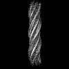
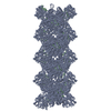
EMDB-50318: 
3D Cryo-EM reveals the structure of a 3-Fmoc zipper motif ensuring the self-assembly of tripeptide nanofiber
Method: helical / : Estrozi LF, Jierry L
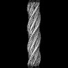
EMDB-50319:
3D Cryo-EM reveals the structure of a 3-Fmoc zipper motif ensuring the self-assembly of tripeptide nanofibers
Method: helical / : Estrozi LF, Jierry L
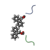
PDB-9fck:
3D Cryo-EM reveals the structure of a 3-Fmoc zipper motif ensuring the self-assembly of tripeptide nanofiber
Method: helical / : Estrozi LF, Jierry L
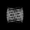
EMDB-19822:
Helical reconstruction of yeast eisosome protein Pil1 bound to membrane composed of lipid mixture +PIP2/+bromosterol (DOPC, DOPE, DOPS, bromo-ergosterol, PI(4,5)P2 35:20:20:15:10)
Method: helical / : Kefauver JM, Zou L, Desfosses A, Loewith RJ
EMDB-18307:
Native eisosome lattice bound to plasma membrane microdomain
Method: single particle / : Kefauver JM, Zou L, Loewith RJ, Desfosses A
EMDB-18308:
Helical reconstruction of yeast eisosome protein Pil1 bound to membrane composed of lipid mixture -PIP2/+sterol (DOPC, DOPE, DOPS, cholesterol 30:20:20:30)
Method: helical / : Kefauver JM, Zou L, Desfosses A, Loewith RJ
EMDB-18309:
Helical reconstruction of yeast eisosome protein Pil1 bound to membrane composed of lipid mixture +PIP2/-sterol (DOPC, DOPE, DOPS, PI(4,5)P2 50:20:20:10)
Method: helical / : Kefauver JM, Zou L, Desfosses A, Loewith RJ
EMDB-18310:
Helical reconstruction of yeast eisosome protein Pil1 bound to membrane composed of lipid mixture +PIP2/+sterol (DOPC, DOPE, DOPS, cholesterol, PI(4,5)P2 35:20:20:15:10)
Method: helical / : Kefauver JM, Zou L, Desfosses A, Loewith RJ
EMDB-18311:
Compact state - Native eisosome lattice bound to plasma membrane microdomain
Method: single particle / : Kefauver JM, Zou L, Desfosses A, Loewith RJ
EMDB-18312:
Stretched state - Native eisosome lattice bound to plasma membrane microdomain
Method: single particle / : Kefauver JM, Zou L, Desfosses A, Loewith RJ
PDB-8qb7:
Pil1 in native eisosome lattice bound to plasma membrane microdomain
Method: single particle / : Kefauver JM, Zou L, Loewith RJ, Desfosses A
PDB-8qb8:
Lsp1 in native eisosome lattice bound to plasma membrane microdomain
Method: single particle / : Kefauver JM, Zou L, Loewith RJ, Desfosses A
PDB-8qb9:
Helical reconstruction of yeast eisosome protein Pil1 bound to membrane composed of lipid mixture -PIP2/+sterol (DOPC, DOPE, DOPS, cholesterol 30:20:20:30)
Method: helical / : Kefauver JM, Zou L, Desfosses A, Loewith RJ
PDB-8qbb:
Helical reconstruction of yeast eisosome protein Pil1 bound to membrane composed of lipid mixture +PIP2/-sterol (DOPC, DOPE, DOPS, PI(4,5)P2 50:20:20:10)
Method: helical / : Kefauver JM, Zou L, Desfosses A, Loewith RJ
PDB-8qbd:
Helical reconstruction of yeast eisosome protein Pil1 bound to membrane composed of lipid mixture +PIP2/+sterol (DOPC, DOPE, DOPS, cholesterol, PI(4,5)P2 35:20:20:15:10)
Method: helical / : Kefauver JM, Zou L, Desfosses A, Loewith RJ
PDB-8qbe:
Compact state - Pil1 in native eisosome lattice bound to plasma membrane microdomain
Method: single particle / : Kefauver JM, Zou L, Desfosses A, Loewith RJ
PDB-8qbf:
Compact state - Pil1 dimer with lipid headgroups fitted in native eisosome lattice bound to plasma membrane microdomain
Method: single particle / : Kefauver JM, Zou L, Desfosses A, Loewith RJ
PDB-8qbg:
Stretched state - Pil1 in native eisosome lattice bound to plasma membrane microdomain
Method: single particle / : Kefauver JM, Zou L, Desfosses A, Loewith RJ
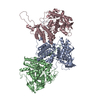
EMDB-18043:
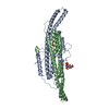
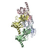
Helical structure of the influenza A virus ribonucleoprotein-like
Method: helical / : Chenavier F, Estrozi LF, Zarkadas E, Ruigrok RWH, Schoehn G, Ballandras-Colas A, Crepin T
EMDB-18044:
Focused reconstruction of influenza A RNP-like particle
Method: helical / : Chenavier F, Estrozi LF, Zarkadas E, Ruigrok RWH, Schoehn G, Ballandras-Colas A, Crepin T
PDB-8pzp:
Model for influenza A virus helical ribonucleoprotein-like structure
Method: helical / : Chenavier F, Estrozi LF, Zarkadas E, Ruigrok RWH, Schoehn G, Ballandras-Colas A, Crepin T
PDB-8pzq:
Model for focused reconstruction of influenza A RNP-like particle
Method: helical / : Chenavier F, Estrozi LF, Zarkadas E, Ruigrok RWH, Schoehn G, Ballandras-Colas A, Crepin T
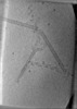
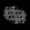
EMDB-15627:
Tomogram of the Mimivirus genomic fiber
Method: electron tomography / : Villalta A, Schmitt A, Estrozi L, Quemin ERK, Alempic JM, Lartigue A, Prazak V, Belmudes L, Vasishtan D, Colmant AMG, Honore FA, Coute Y, Gruenewald K, Abergel C
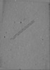
EMDB-15628:
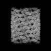
Tomogram of the Mimivirus genomic fiber
Method: electron tomography / : Villalta A, Schmitt A, Estrozi L, Quemin ERK, Alempic JM, Lartigue A, Prazak V, Belmudes L, Vasishtan D, Colmant AMG, Honore FA, Coute Y, Gruenewald K, Abergel C
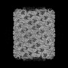
EMDB-15629:
Tomogram of the Mimivirus genomic fiber
Method: electron tomography / : Villalta A, Schmitt A, Estrozi L, Quemin ERK, Alempic JM, Lartigue A, Prazak V, Belmudes L, Vasishtan D, Colmant AMG, Honore FA, Coute Y, Gruenewald K, Abergel C
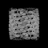
EMDB-15630:
Tomogram of the Mimivirus genomic fiber
Method: electron tomography / : Villalta A, Schmitt A, Estrozi L, Quemin ERK, Alempic JM, Lartigue A, Prazak V, Belmudes L, Vasishtan D, Colmant AMG, Honore FA, Coute Y, Gruenewald K, Abergel C

EMDB-13641:
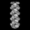
Structure of the Mimivirus genomic fibre asymmetric unit
Method: single particle / : Villalta A, Schmitt A, Estrozi LF, Quemin ERJ, Alempic JM, Lartigue A, Prazak V, Belmudes L, Vasishtan D, Colmant AMG, Honore FA, Coute Y, Grunewald K, Abergel C

EMDB-14353:
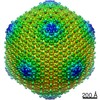
Structure of the Mimivirus genomic fibre in its compact 6-start helix form
Method: helical / : Villalta A, Schmitt A

EMDB-14354:
Structure of the Mimivirus genomic fibre in its compact 5-start helix form
Method: helical / : Villalta A, Schmitt A

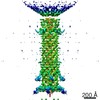
EMDB-14355:
Structure of the Mimivirus genomic fibre in its relaxed 5-start helix form
Method: helical / : Villalta A, Schmitt A
PDB-7ptv:
Structure of the Mimivirus genomic fibre asymmetric unit
Method: single particle / : Villalta A, Schmitt A, Estrozi LF, Quemin ERJ, Alempic JM, Lartigue A, Prazak V, Belmudes L, Vasishtan D, Colmant AMG, Honore FA, Coute Y, Grunewald K, Abergel C
PDB-7yx3:
Structure of the Mimivirus genomic fibre in its compact 6-start helix form
Method: helical / : Villalta A, Schmitt A, Estrozi LF, Quemin ERJ, Alempic JM, Lartigue A, Prazak V, Belmudes L, Vasishtan D, Colmant AMG, Honore FA, Coute Y, Grunewald K, Abergel C
PDB-7yx4:
Structure of the Mimivirus genomic fibre in its compact 5-start helix form
Method: helical / : Villalta A, Schmitt A, Estrozi LF, Quemin ERJ, Alempic JM, Lartigue A, Prazak V, Belmudes L, Vasishtan D, Colmant AMG, Honore FA, Coute Y, Grunewald K, Abergel C
PDB-7yx5:
Structure of the Mimivirus genomic fibre in its relaxed 5-start helix form
Method: helical / : Villalta A, Schmitt A, Estrozi LF, Quemin ERJ, Alempic JM, Lartigue A, Prazak V, Belmudes L, Vasishtan D, Colmant AMG, Honore FA, Coute Y, Grunewald K, Abergel C
EMDB-11179:
Head of Semi-jumbo phage RP13
Method: single particle / : Neumann E, Kawasaki T, Effantin G, Estrozi L, Chatchawankanphanich O, Yamada T, Schoehn G
EMDB-11180:
Head reconstruction of full jumbo phage XacN1
Method: single particle / : Neumann E, Kawasaki T, Effantin G, Estrozi L, Chatchawankanphanich O, Yamada T, Schoehn G

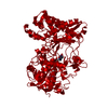
EMDB-11275:
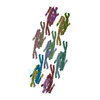
MreC
Method: helical / : Estrozi LF, Contreras-Martel C
PDB-6zlv:
MreC
Method: helical / : Estrozi LF, Contreras-Martel C
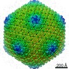
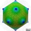
EMDB-11178:
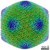
Jumbo Bacteriophage RSL2 - Full icosahedral capsid
Method: single particle / : Neumann E, Kawasaki T, Effantin G, Estrozi L, Chatchawankanphanich O, Yamada T, Schoehn G
EMDB-10926:
Structure of jumbo coliphage phAPEC6 capsid
Method: single particle / : Wagemans J, Tsonos J, Holtappels D, Fortuna K, Hernalsteens JP, De Greve H, Estrozi LF, Bacia-Verloop M, Moriscot C, Noben JP, Schoehn G, Lavigne R
EMDB-10929:
3D structure of bacteriophage phAPEC6 tail
Method: single particle / : Wagemans J, Tsonos J, Holtappels D, Fortuna K, Hernalsteens JP, De Greve H, Estrozi LF, Bacia-Verloop M, Moriscot C, Noben JP, Schoehn G, Lavigne R
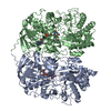
PDB-6r8n:
STRUCTURE DETERMINATION OF THE TETRAHEDRAL AMINOPEPTIDASE TET2 FROM P. HORIKOSHII BY USE OF COMBINED SOLID-STATE NMR, SOLUTION-STATE NMR AND EM DATA 4.1 A, FOLLOWED BY REAL_SPACE_REFINEMENT AT 4.1 A
Method: single particle / : Colletier JP, Gauto D, Estrozi L, Favier A, Effantin G, Schoehn G, Boisbouvier J, Schanda P
EMDB-20086:
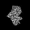
In situ structure of rotavirus VP1 RNA-dependent RNA polymerase (TLP)
Method: single particle / : Jenni S, Salgado EN
EMDB-20087:
In situ structure of rotavirus VP1 RNA-dependent RNA polymerase (DLP)
Method: single particle / : Jenni S, Salgado EN
EMDB-20088:
In situ structure of rotavirus VP1 RNA-dependent RNA polymerase (TLP_RNA)
Method: single particle / : Jenni S, Salgado EN

EMDB-20089:
In situ structure of rotavirus VP1 RNA-dependent RNA polymerase (DLP_RNA)
Method: single particle / : Jenni S, Salgado EN
PDB-6oj3:
In situ structure of rotavirus VP1 RNA-dependent RNA polymerase (TLP)
Method: single particle / : Jenni S, Salgado EN, Herrmann T, Li Z, Grant T, Grigorieff N, Trapani S, Estrozi LF, Harrison SC
PDB-6oj4:
In situ structure of rotavirus VP1 RNA-dependent RNA polymerase (DLP)
Method: single particle / : Jenni S, Salgado EN, Herrmann T, Li Z, Grant T, Grigorieff N, Trapani S, Estrozi LF, Harrison SC
PDB-6oj5:
In situ structure of rotavirus VP1 RNA-dependent RNA polymerase (TLP_RNA)
Method: single particle / : Jenni S, Salgado EN, Herrmann T, Li Z, Grant T, Grigorieff N, Trapani S, Estrozi LF, Harrison SC
PDB-6oj6:
In situ structure of rotavirus VP1 RNA-dependent RNA polymerase (DLP_RNA)
Method: single particle / : Jenni S, Salgado EN, Herrmann T, Li Z, Grant T, Grigorieff N, Trapani S, Estrozi LF, Harrison SC
Pages:
 wwPDB to switch to version 3 of the EMDB data model
