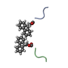
Journal: ACS Nano / Year: 2024 Title: 3D Cryo-Electron Microscopy Reveals the Structure of a 3-Fluorenylmethyloxycarbonyl Zipper Motif Ensuring the Self-Assembly of Tripeptide Nanofibers. Authors: Alexis Bigo-Simon / Leandro F Estrozi / Alain Chaumont / Rachel Schurhammer / Guy Schoehn / Jérôme Combet / Marc Schmutz / Pierre Schaaf / Loïc Jierry / Abstract: Short peptide-based supramolecular hydrogels appeared as highly interesting materials for applications in many fields. The optimization of their properties relies mainly on the design of a suitable ...Short peptide-based supramolecular hydrogels appeared as highly interesting materials for applications in many fields. The optimization of their properties relies mainly on the design of a suitable hydrogelator through an empirical trial-and-error strategy based on the synthesis of various types of peptides. This approach is in part due to the lack of prior structural knowledge of the molecular architecture of the various families of nanofibers. The 3D structure of the nanofibers determines their ability to interact with entities present in their surrounding environment. Thus, it is important to resolve the internal structural organization of the material. Herein, using Fmoc-FFY tripeptide as a model amphiphilic hydrogelator and cryo-EM reconstruction approach, we succeeded to obtain a 3.8 Å resolution 3D structure of a self-assembled nanofiber with a diameter of approximately 4.1 nm and with apparently "infinite" length. The elucidation of the spatial organization of such nano-objects addresses fundamental questions about the way short amphiphilic -Fmoc peptides lacking secondary structure can self-assemble and ensure the cohesion of such a lengthy nanostructure. This nanofiber is organized into a triple-stranded helix with an asymmetric unit composed of two Fmoc-FFY peptides per strand. The three identical amphiphilic strands are maintained together by strong lateral interactions coming from a 3-Fmoc zipper motif. This hydrophobic core of the nanofiber is surrounded by 12 phenyl groups from phenylalanine residues, nonplanar with the six Fmoc groups. Polar tyrosine residues at the C-term position constitute the hydrophilic shell and are exposed all around the external part of the assembly. This fiber has a highly hydrophobic central core with an internal diameter of only 2.4 Å. Molecular dynamics simulations highlight van der Waals and hydrogen bonds between peptides placed on top of each other. We demonstrate that the self-assembly of Fmoc-FFY, whether induced by annealing or by the action of a phosphatase on the phosphorylated precursor Fmoc-FFY, results in two nanostructures with minor differences that we are unable to distinguish.
In the structure databanks used in Yorodumi, some data are registered as the other names, "COVID-19 virus" and "2019-nCoV". Here are the details of the virus and the list of structure data.
Jan 31, 2019. EMDB accession codes are about to change! (news from PDBe EMDB page)
EMDB accession codes are about to change! (news from PDBe EMDB page)
The allocation of 4 digits for EMDB accession codes will soon come to an end. Whilst these codes will remain in use, new EMDB accession codes will include an additional digit and will expand incrementally as the available range of codes is exhausted. The current 4-digit format prefixed with “EMD-” (i.e. EMD-XXXX) will advance to a 5-digit format (i.e. EMD-XXXXX), and so on. It is currently estimated that the 4-digit codes will be depleted around Spring 2019, at which point the 5-digit format will come into force.
The EM Navigator/Yorodumi systems omit the EMD- prefix.
Related info.:Q: What is EMD? / ID/Accession-code notation in Yorodumi/EM Navigator
Yorodumi is a browser for structure data from EMDB, PDB, SASBDB, etc.
This page is also the successor to EM Navigator detail page, and also detail information page/front-end page for Omokage search.
The word "yorodu" (or yorozu) is an old Japanese word meaning "ten thousand". "mi" (miru) is to see.
Related info.:EMDB / PDB / SASBDB / Comparison of 3 databanks / Yorodumi Search / Aug 31, 2016. New EM Navigator & Yorodumi / Yorodumi Papers / Jmol/JSmol / Function and homology information / Changes in new EM Navigator and Yorodumi
 Movie
Movie Controller

 Yorodumi
 Open data
 Basic information
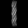
 Map data
 Sample
 Keywords
 Authors
 France, 1 items
  Citation
 Structure visualization
 Downloads & links
 EMDB map data format
 emd_50318.png
 http://ftp.pdbj.org/pub/emdb/structures/EMD-50318
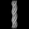
 Z (Sec.)
 Y (Row.)
 X (Col.)


 Sample components
 Processing
 Electron microscopy
 FIELD EMISSION GUN