 Movie
Movie Controller

[English] 日本語
 Yorodumi
- PDB-6r8n: STRUCTURE DETERMINATION OF THE TETRAHEDRAL AMINOPEPTIDASE TET2 FR... -
+ Open data

- Basic information
| Entry | Database: PDB / ID: 6r8n | |||||||||||||||||||||
|---|---|---|---|---|---|---|---|---|---|---|---|---|---|---|---|---|---|---|---|---|---|---|
| Title | STRUCTURE DETERMINATION OF THE TETRAHEDRAL AMINOPEPTIDASE TET2 FROM P. HORIKOSHII BY USE OF COMBINED SOLID-STATE NMR, SOLUTION-STATE NMR AND EM DATA 4.1 A, FOLLOWED BY REAL_SPACE_REFINEMENT AT 4.1 A | |||||||||||||||||||||
 Components Components | Tetrahedral aminopeptidase | |||||||||||||||||||||
 Keywords Keywords | PEPTIDE BINDING PROTEIN / PEPTIDASE / PROTEIN QUALITY CONTROL / OLIGOMER / AMINOPEPTIDASE | |||||||||||||||||||||
| Function / homology |  Function and homology information Function and homology informationHydrolases; Acting on peptide bonds (peptidases); Aminopeptidases / aminopeptidase activity / metallopeptidase activity / proteolysis / metal ion binding Similarity search - Function | |||||||||||||||||||||
| Biological species |   Pyrococcus horikoshii OT3 (archaea) Pyrococcus horikoshii OT3 (archaea) | |||||||||||||||||||||
| Method | ELECTRON MICROSCOPY / SOLUTION NMR / single particle reconstruction / simulated annealing / cryo EM / Resolution: 4.1 Å | |||||||||||||||||||||
 Authors Authors | Colletier, J.-P. / Gauto, D. / Estrozi, L. / Favier, A. / Effantin, G. / Schoehn, G. / Boisbouvier, J. / Schanda, P. | |||||||||||||||||||||
| Funding support |  France, 6items France, 6items
| |||||||||||||||||||||
 Citation Citation | Journal: Nat Commun / Year: 2019 Title: Integrated NMR and cryo-EM atomic-resolution structure determination of a half-megadalton enzyme complex. Authors: Diego F Gauto / Leandro F Estrozi / Charles D Schwieters / Gregory Effantin / Pavel Macek / Remy Sounier / Astrid C Sivertsen / Elena Schmidt / Rime Kerfah / Guillaume Mas / Jacques-Philippe ...Authors: Diego F Gauto / Leandro F Estrozi / Charles D Schwieters / Gregory Effantin / Pavel Macek / Remy Sounier / Astrid C Sivertsen / Elena Schmidt / Rime Kerfah / Guillaume Mas / Jacques-Philippe Colletier / Peter Güntert / Adrien Favier / Guy Schoehn / Paul Schanda / Jerome Boisbouvier /     Abstract: Atomic-resolution structure determination is crucial for understanding protein function. Cryo-EM and NMR spectroscopy both provide structural information, but currently cryo-EM does not routinely ...Atomic-resolution structure determination is crucial for understanding protein function. Cryo-EM and NMR spectroscopy both provide structural information, but currently cryo-EM does not routinely give access to atomic-level structural data, and, generally, NMR structure determination is restricted to small (<30 kDa) proteins. We introduce an integrated structure determination approach that simultaneously uses NMR and EM data to overcome the limits of each of these methods. The approach enables structure determination of the 468 kDa large dodecameric aminopeptidase TET2 to a precision and accuracy below 1 Å by combining secondary-structure information obtained from near-complete magic-angle-spinning NMR assignments of the 39 kDa-large subunits, distance restraints from backbone amides and ILV methyl groups, and a 4.1 Å resolution EM map. The resulting structure exceeds current standards of NMR and EM structure determination in terms of molecular weight and precision. Importantly, the approach is successful even in cases where only medium-resolution cryo-EM data are available. | |||||||||||||||||||||
| History |
|
- Structure visualization
| Movie |
Movie viewer |
|---|---|
| Structure viewer | Molecule: MolmilJmol/JSmol |
- Downloads & links
-Download
| PDBx/mmCIF format | 6r8n.cif.gz | 1.3 MB | Display | PDBx/mmCIF format |
|---|---|---|---|---|
| PDB format | pdb6r8n.ent.gz | 1.1 MB | Display | PDB format |
| PDBx/mmJSON format | 6r8n.json.gz | Tree view | PDBx/mmJSON format | |
| Others |  Other downloads Other downloads |
-Validation report
| Arichive directory | https://data.pdbj.org/pub/pdb/validation_reports/r8/6r8nftp://data.pdbj.org/pub/pdb/validation_reports/r8/6r8n | HTTPS FTP |
|---|
-Related structure data
| Related structure data | 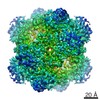 4179MC  6f3kC M: map data used to model this data C: citing same article ( |
|---|---|
| Similar structure data | |
| Other databases |
|

-Links
 PDBj
PDBj- Assembly
| Deposited unit | 
| |||||||||
|---|---|---|---|---|---|---|---|---|---|---|
| 1 |
| |||||||||
| NMR ensembles |
|
-Components
| #1: Protein | Mass: 39071.027 Da / Num. of mol.: 12 Source method: isolated from a genetically manipulated source Source: (gene. exp.) Pyrococcus horikoshii OT3 (archaea) / Gene: frvX, PH1527Variant: ATCC 700860 / DSM 12428 / JCM 9974 / NBRC 100139 / OT-3 Production host:  References: UniProt: O59196, Hydrolases; Acting on peptide bonds (peptidases); Aminopeptidases #2: Chemical | ChemComp-ZN /   Mass: 65.409 Da / Num. of mol.: 24 / Source method: obtained synthetically / Formula: Zn Mass: 65.409 Da / Num. of mol.: 24 / Source method: obtained synthetically / Formula: Zn |
|---|
-Experimental details
-Experiment
| Experiment |
| ||||||||||||||||||||||||||||||||||||||||||||||||||||||||||||||||||||||||||||||
|---|---|---|---|---|---|---|---|---|---|---|---|---|---|---|---|---|---|---|---|---|---|---|---|---|---|---|---|---|---|---|---|---|---|---|---|---|---|---|---|---|---|---|---|---|---|---|---|---|---|---|---|---|---|---|---|---|---|---|---|---|---|---|---|---|---|---|---|---|---|---|---|---|---|---|---|---|---|---|---|
| EM experiment | Aggregation state: PARTICLE / 3D reconstruction method: single particle reconstruction | ||||||||||||||||||||||||||||||||||||||||||||||||||||||||||||||||||||||||||||||
| NMR experiment |
|
- Sample preparation
| Component | Name: TETRAHEDRAL AMINOPEPTIDASE TET2 / Type: COMPLEX Details: The structure was built using combined SOLID-STATE NMR, SOLUTION-STATE NMR AND EM DATA, and then refined using phenix.real_space_refine Entity ID: #1 / Source: RECOMBINANT | ||||||||||||||||||||||||||||||||||||||||||||
|---|---|---|---|---|---|---|---|---|---|---|---|---|---|---|---|---|---|---|---|---|---|---|---|---|---|---|---|---|---|---|---|---|---|---|---|---|---|---|---|---|---|---|---|---|---|
| Molecular weight | Experimental value: NO | ||||||||||||||||||||||||||||||||||||||||||||
| Source (natural) | Organism: Pyrococcus horikoshii OT3 (archaea) | ||||||||||||||||||||||||||||||||||||||||||||
| Source (recombinant) | Organism: | ||||||||||||||||||||||||||||||||||||||||||||
| Buffer solution | pH: 7.4 | ||||||||||||||||||||||||||||||||||||||||||||
| Specimen | Conc.: 1 mg/ml / Embedding applied: NO / Shadowing applied: NO / Staining applied: NO / Vitrification applied: YES / Details: This sample was monodisperse | ||||||||||||||||||||||||||||||||||||||||||||
| Specimen support | Grid material: COPPER / Grid mesh size: 400 divisions/in. / Grid type: Quantifoil, UltrAuFoil, R1.2/1.3 | ||||||||||||||||||||||||||||||||||||||||||||
| Vitrification | Instrument: FEI VITROBOT MARK IV / Cryogen name: ETHANE / Humidity: 100 % / Chamber temperature: 293 K / Details: Blotting time 2s, force 1, drain time 0. | ||||||||||||||||||||||||||||||||||||||||||||
| Details |
| ||||||||||||||||||||||||||||||||||||||||||||
| Sample |
| ||||||||||||||||||||||||||||||||||||||||||||
| Sample conditions |
|
-Data collection
| Experimental equipment |  Model: Tecnai Polara / Image courtesy: FEI Company | ||||||||||||||||||||
|---|---|---|---|---|---|---|---|---|---|---|---|---|---|---|---|---|---|---|---|---|---|
| Microscopy | Model: FEI POLARA 300 | ||||||||||||||||||||
| Electron gun | Electron source:  FIELD EMISSION GUN / Accelerating voltage: 300 kV / Illumination mode: FLOOD BEAM FIELD EMISSION GUN / Accelerating voltage: 300 kV / Illumination mode: FLOOD BEAM | ||||||||||||||||||||
| Electron lens | Mode: BRIGHT FIELD / Nominal magnification: 20000 X / Calibrated magnification: 27773 X / Calibrated defocus min: 8000 nm / Calibrated defocus max: 30000 nm / Cs: 2 mm / Alignment procedure: COMA FREE | ||||||||||||||||||||
| Specimen holder | Cryogen: NITROGEN Specimen holder model: GATAN 626 SINGLE TILT LIQUID NITROGEN CRYO TRANSFER HOLDER | ||||||||||||||||||||
| Image recording | Average exposure time: 4 sec. / Electron dose: 40 e/Å2 / Detector mode: SUPER-RESOLUTION / Film or detector model: GATAN K2 SUMMIT (4k x 4k) / Num. of real images: 90 | ||||||||||||||||||||
| Image scans | Movie frames/image: 40 | ||||||||||||||||||||
| NMR spectrometer |
|
- Processing
| Software | Name: PHENIX / Version: 1.14_3260: / Classification: refinement | ||||||||||||||||||||||||||||||||||||
|---|---|---|---|---|---|---|---|---|---|---|---|---|---|---|---|---|---|---|---|---|---|---|---|---|---|---|---|---|---|---|---|---|---|---|---|---|---|
| EM software |
| ||||||||||||||||||||||||||||||||||||
| CTF correction | Type: PHASE FLIPPING AND AMPLITUDE CORRECTION | ||||||||||||||||||||||||||||||||||||
| Particle selection | Num. of particles selected: 30407 | ||||||||||||||||||||||||||||||||||||
| Symmetry | Point symmetry: T (tetrahedral) | ||||||||||||||||||||||||||||||||||||
| 3D reconstruction | Resolution: 4.1 Å / Resolution method: FSC 0.143 CUT-OFF / Num. of particles: 27130 / Algorithm: FOURIER SPACE / Symmetry type: POINT | ||||||||||||||||||||||||||||||||||||
| Atomic model building | Protocol: OTHER | ||||||||||||||||||||||||||||||||||||
| NMR software |
| ||||||||||||||||||||||||||||||||||||
| Refinement | Method: simulated annealing / Software ordinal: 7 | ||||||||||||||||||||||||||||||||||||
| NMR representative | Selection criteria: lowest energy | ||||||||||||||||||||||||||||||||||||
| NMR ensemble | Conformer selection criteria: structures with the lowest energy Conformers calculated total number: 100 / Conformers submitted total number: 1 |
