 Movie
Movie Controller


+ Open data

- Basic information
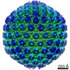
| Entry | Database: EMDB / ID: EMD-11178 | |||||||||
|---|---|---|---|---|---|---|---|---|---|---|
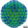
| Title | Jumbo Bacteriophage RSL2 - Full icosahedral capsid | |||||||||
 Map data Map data | Head Reconstruction of the jumbo phage RSL2 | |||||||||
 Sample Sample |
| |||||||||
| Biological species |  Ralstonia virus RSL2 Ralstonia virus RSL2 | |||||||||
| Method | single particle reconstruction / cryo EM / Resolution: 16.0 Å | |||||||||
 Authors Authors | Neumann E / Kawasaki T / Effantin G / Estrozi L / Chatchawankanphanich O / Yamada T / Schoehn G | |||||||||
 Citation Citation | Journal: J Gen Virol / Year: 2020 Title: 3D structure of three jumbo phage heads. Authors: Emmanuelle Neumann / Takeru Kawasaki / Grégory Effantin / Leandro F Estrozi / Orawan Chatchawankanphanich / Takashi Yamada / Guy Schoehn /    Abstract: Jumbo phages are bacteriophages that carry more than 200 kbp of DNA. In this study we characterized two jumbo phages (ΦRSL2 and ΦXacN1) and one semi-jumbo phage (ΦRP13) at the structural level by ...Jumbo phages are bacteriophages that carry more than 200 kbp of DNA. In this study we characterized two jumbo phages (ΦRSL2 and ΦXacN1) and one semi-jumbo phage (ΦRP13) at the structural level by cryo-electron microscopy. Focusing on their capsids, three-dimensional structures of the heads at resolutions ranging from 16 to 9 Å were calculated. Based on these structures we determined the geometrical basis on which the icosahedral capsids of these phages are constructed, which includes the accessory and decorative proteins that complement them. A triangulation number novel to (ΦRP13; =21) was discovered as well as two others, which are more common for jumbo phages (=27 and =28). Based on one of the structures we also provide evidence that accessory or decorative proteins are not a prerequisite for maintaining the structural integrity of very large capsids. | |||||||||
| History |
|
- Structure visualization
| Movie |
 Movie viewer Movie viewer |
|---|---|
| Structure viewer | EM map: SurfViewMolmilJmol/JSmol |
| Supplemental images |

- Downloads & links
-EMDB archive
| Map data | emd_11178.map.gz | 48.5 MB | EMDB map data format | |
|---|---|---|---|---|
| Header (meta data) | emd-11178-v30.xmlemd-11178.xml | 9.9 KB 9.9 KB | Display Display | EMDB header |
| FSC (resolution estimation) | emd_11178_fsc.xml | 11 KB | Display | FSC data file |
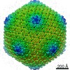
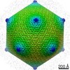
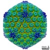
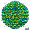
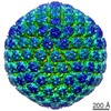
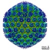
| Images | 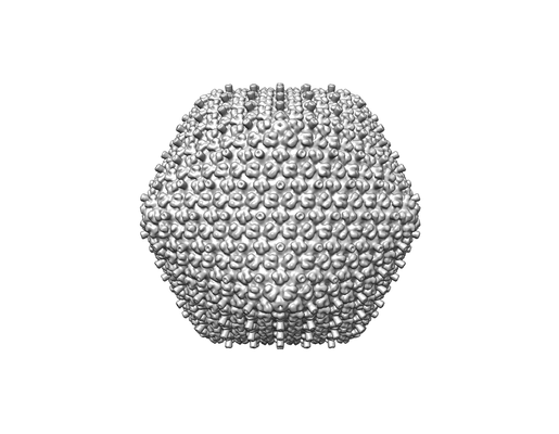 emd_11178.png emd_11178.png | 50.2 KB | ||
| Archive directory |  http://ftp.pdbj.org/pub/emdb/structures/EMD-11178ftp://ftp.pdbj.org/pub/emdb/structures/EMD-11178 http://ftp.pdbj.org/pub/emdb/structures/EMD-11178ftp://ftp.pdbj.org/pub/emdb/structures/EMD-11178 | HTTPS FTP |
-Related structure data
| Related structure data | C: citing same article ( |
|---|---|
| Similar structure data |
-Links
| EMDB pages | EMDB (EBI/PDBe) / EMDataResource |
|---|
-Map
| File | Download / File: emd_11178.map.gz / Format: CCP4 / Size: 52.7 MB / Type: IMAGE STORED AS FLOATING POINT NUMBER (4 BYTES) | ||||||||||||||||||||||||||||||||||||||||||||||||||||||||||||
|---|---|---|---|---|---|---|---|---|---|---|---|---|---|---|---|---|---|---|---|---|---|---|---|---|---|---|---|---|---|---|---|---|---|---|---|---|---|---|---|---|---|---|---|---|---|---|---|---|---|---|---|---|---|---|---|---|---|---|---|---|---|
| Annotation | Head Reconstruction of the jumbo phage RSL2 | ||||||||||||||||||||||||||||||||||||||||||||||||||||||||||||
| Projections & slices | Image control
Images are generated by Spider. | ||||||||||||||||||||||||||||||||||||||||||||||||||||||||||||
| Voxel size | X=Y=Z: 6.57 Å | ||||||||||||||||||||||||||||||||||||||||||||||||||||||||||||
| Density |
| ||||||||||||||||||||||||||||||||||||||||||||||||||||||||||||
| Symmetry | Space group: 1 | ||||||||||||||||||||||||||||||||||||||||||||||||||||||||||||
| Details | EMDB XML:
CCP4 map header:
| ||||||||||||||||||||||||||||||||||||||||||||||||||||||||||||
 Z (Sec.)
 Y (Row.)
 X (Col.)


-Supplemental data
- Sample components
-Entire : Ralstonia virus RSL2
| Entire | Name: Ralstonia virus RSL2 |
|---|---|
| Components |
|
-Supramolecule #1: Ralstonia virus RSL2
| Supramolecule | Name: Ralstonia virus RSL2 / type: virus / ID: 1 / Parent: 0 / NCBI-ID: 1980925 / Sci species name: Ralstonia virus RSL2 / Virus type: VIRION / Virus isolate: OTHER / Virus enveloped: No / Virus empty: No |
|---|---|
| Host (natural) | Organism: Ralstonia solanacearum MAFF 106603 |
| Virus shell | Shell ID: 1 / Name: RSL2 Head / Diameter: 1390.0 Å / T number (triangulation number): 27 |
-Experimental details
-Structure determination
| Method | cryo EM |
|---|---|
 Processing Processing | single particle reconstruction |
| Aggregation state | particle |
-Sample preparation
| Buffer | pH: 7.5 |
|---|---|
| Grid | Model: Quantifoil / Material: COPPER / Mesh: 400 / Pretreatment - Type: GLOW DISCHARGE |
| Vitrification | Cryogen name: ETHANE / Chamber humidity: 100 % / Chamber temperature: 293 K / Instrument: FEI VITROBOT MARK IV |
- Electron microscopy
| Microscope | FEI POLARA 300 |
|---|---|
| Image recording | Film or detector model: GATAN K2 SUMMIT (4k x 4k) / Detector mode: COUNTING / Average electron dose: 40.0 e/Å2 |
| Electron beam | Acceleration voltage: 300 kV / Electron source:  FIELD EMISSION GUN FIELD EMISSION GUN |
| Electron optics | Illumination mode: OTHER / Imaging mode: BRIGHT FIELD / Cs: 2.0 mm |
| Experimental equipment |  Model: Tecnai Polara / Image courtesy: FEI Company |
+Image processing

-Atomic model buiding 1
| Refinement | Protocol: AB INITIO MODEL |
|---|
