 Movie
Movie Controller


+ Open data

- Basic information
| Entry | Database: EMDB / ID: EMD-10926 | |||||||||
|---|---|---|---|---|---|---|---|---|---|---|
| Title | Structure of jumbo coliphage phAPEC6 capsid | |||||||||
 Map data Map data | Bacteriophage phAPEC6 capsid at 9A resolution | |||||||||
 Sample Sample |
| |||||||||
| Biological species |  | |||||||||
| Method | single particle reconstruction / cryo EM / Resolution: 10.0 Å | |||||||||
 Authors Authors | Wagemans J / Tsonos J / Holtappels D / Fortuna K / Hernalsteens JP / De Greve H / Estrozi LF / Bacia-Verloop M / Moriscot C / Noben JP ...Wagemans J / Tsonos J / Holtappels D / Fortuna K / Hernalsteens JP / De Greve H / Estrozi LF / Bacia-Verloop M / Moriscot C / Noben JP / Schoehn G / Lavigne R | |||||||||
 Citation Citation | Journal: Int J Mol Sci / Year: 2020 Title: Structural Analysis of Jumbo Coliphage phAPEC6. Authors: Jeroen Wagemans / Jessica Tsonos / Dominique Holtappels / Kiandro Fortuna / Jean-Pierre Hernalsteens / Henri De Greve / Leandro F Estrozi / Maria Bacia-Verloop / Christine Moriscot / Jean- ...Authors: Jeroen Wagemans / Jessica Tsonos / Dominique Holtappels / Kiandro Fortuna / Jean-Pierre Hernalsteens / Henri De Greve / Leandro F Estrozi / Maria Bacia-Verloop / Christine Moriscot / Jean-Paul Noben / Guy Schoehn / Rob Lavigne /   Abstract: The phAPEC6 genome encodes 551 predicted gene products, with the vast majority (83%) of unknown function. Of these, 62 have been identified as virion-associated proteins by mass spectrometry (ESI- ...The phAPEC6 genome encodes 551 predicted gene products, with the vast majority (83%) of unknown function. Of these, 62 have been identified as virion-associated proteins by mass spectrometry (ESI-MS/MS), including the major capsid protein (Gp225; present in 1620 copies), which shows a HK97 capsid protein-based fold. Cryo-electron microscopy experiments showed that the 350-kbp DNA molecule of virus phAPEC6 is packaged in at least 15 concentric layers in the phage capsid. A capsid inner body rod is also present, measuring about 91 nm by 18 nm and oriented along the portal axis. In the phAPEC6 contractile tail, 25 hexameric stacked rings can be distinguished, built of the identified tail sheath protein (Gp277). Cryo-EM reconstruction reveals the base of the unique hairy fibers observed during an initial transmission electron microscopy (TEM) analysis. These very unusual filaments are ordered at three annular positions along the contractile sheath, as well as around the capsid, and may be involved in host interaction. | |||||||||
| History |
|
- Structure visualization
| Movie |
 Movie viewer Movie viewer |
|---|---|
| Structure viewer | EM map: SurfViewMolmilJmol/JSmol |
| Supplemental images |

- Downloads & links
-EMDB archive
| Map data | emd_10926.map.gz | 153.8 MB | EMDB map data format | |
|---|---|---|---|---|
| Header (meta data) | emd-10926-v30.xmlemd-10926.xml | 9.1 KB 9.1 KB | Display Display | EMDB header |
| Images | 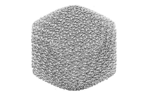 emd_10926.png emd_10926.png | 105.5 KB | ||
| Archive directory |  http://ftp.pdbj.org/pub/emdb/structures/EMD-10926ftp://ftp.pdbj.org/pub/emdb/structures/EMD-10926 http://ftp.pdbj.org/pub/emdb/structures/EMD-10926ftp://ftp.pdbj.org/pub/emdb/structures/EMD-10926 | HTTPS FTP |
-Related structure data
| Related structure data | C: citing same article ( |
|---|---|
| Similar structure data |

-Links
| EMDB pages | EMDB (EBI/PDBe) / EMDataResource |
|---|
-Map
| File | Download / File: emd_10926.map.gz / Format: CCP4 / Size: 515.8 MB / Type: IMAGE STORED AS FLOATING POINT NUMBER (4 BYTES) | ||||||||||||||||||||||||||||||||||||||||||||||||||||||||||||
|---|---|---|---|---|---|---|---|---|---|---|---|---|---|---|---|---|---|---|---|---|---|---|---|---|---|---|---|---|---|---|---|---|---|---|---|---|---|---|---|---|---|---|---|---|---|---|---|---|---|---|---|---|---|---|---|---|---|---|---|---|---|
| Annotation | Bacteriophage phAPEC6 capsid at 9A resolution | ||||||||||||||||||||||||||||||||||||||||||||||||||||||||||||
| Projections & slices | Image control
Images are generated by Spider. generated in cubic-lattice coordinate | ||||||||||||||||||||||||||||||||||||||||||||||||||||||||||||
| Voxel size | X=Y=Z: 2.26 Å | ||||||||||||||||||||||||||||||||||||||||||||||||||||||||||||
| Density |
| ||||||||||||||||||||||||||||||||||||||||||||||||||||||||||||
| Symmetry | Space group: 1 | ||||||||||||||||||||||||||||||||||||||||||||||||||||||||||||
| Details | EMDB XML:
CCP4 map header:
| ||||||||||||||||||||||||||||||||||||||||||||||||||||||||||||
 Z (Sec.)
 Y (Row.)
 X (Col.)


-Supplemental data
- Sample components
-Entire : Escherichia coli
| Entire | Name: |
|---|---|
| Components |
|
-Supramolecule #1: Escherichia coli
| Supramolecule | Name: Escherichia coli / type: virus / ID: 1 / Parent: 0 / NCBI-ID: 562 / Sci species name: Escherichia coli / Virus type: VIRION / Virus isolate: OTHER / Virus enveloped: No / Virus empty: No |
|---|---|
| Virus shell | Shell ID: 1 / Diameter: 1360.0 Å / T number (triangulation number): 28 |
-Experimental details
-Structure determination
| Method | cryo EM |
|---|---|
 Processing Processing | single particle reconstruction |
| Aggregation state | particle |
-Sample preparation
| Concentration | 1 mg/mL |
|---|---|
| Buffer | pH: 7.5 |
| Grid | Model: Quantifoil R2/1 / Material: COPPER / Mesh: 400 / Support film - Material: CARBON / Support film - topology: HOLEY ARRAY / Pretreatment - Type: GLOW DISCHARGE |
| Vitrification | Cryogen name: ETHANE / Chamber humidity: 100 % / Instrument: FEI VITROBOT MARK IV / Details: Force 1, 2s blotting time. |
- Electron microscopy
| Microscope | FEI POLARA 300 |
|---|---|
| Image recording | Film or detector model: KODAK SO-163 FILM / Number grids imaged: 1 / Number real images: 100 / Average electron dose: 20.0 e/Å2 |
| Electron beam | Acceleration voltage: 300 kV / Electron source:  FIELD EMISSION GUN FIELD EMISSION GUN |
| Electron optics | Illumination mode: FLOOD BEAM / Imaging mode: BRIGHT FIELD |
| Experimental equipment |  Model: Tecnai Polara / Image courtesy: FEI Company |
-Image processing
| Particle selection | Number selected: 6900 |
|---|---|
| Final reconstruction | Applied symmetry - Point group: I (icosahedral) / Resolution.type: BY AUTHOR / Resolution: 10.0 Å / Resolution method: FSC 0.143 CUT-OFF / Number images used: 4149 |
| Initial angle assignment | Type: PROJECTION MATCHING |
| Final angle assignment | Type: PROJECTION MATCHING |
