 ムービー
ムービー コントローラー
 構造ビューア
 EMN検索について


-検索条件
-検索結果
検索 (著者・登録者: dutta & m)の結果74件中、1から50件目までを表示しています

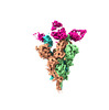
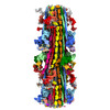
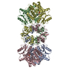
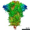
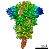
EMDB-36815: 
Recognition determinants of broad and potent HIV-1 neutralization by an affinity matured antibody from a pediatric elite-neutralizer
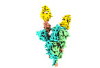
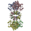
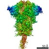
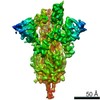
EMDB-34548:
Spike 3-up RBD with THSC20.HVTR04 (Fab4): State - III
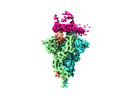
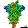
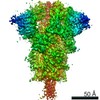
EMDB-34563:
Spike 2-up RBD with THSC20.HVTR26 (Fab26): State - I
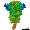
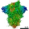
EMDB-34602:
State - I: Spike 2-up RBD with THSC20.HVTR26 (Fab26)

EMDB-34603:
State - II: Spike 3-up RBD with THSC20.HVTR26 (Fab26)

EMDB-34546:
State - I: Spike 2-up RBD with THSC20.HVTR04 (Fab4)
EMDB-34547:
Spike 3-up RBD with THSC20.HVTR04 (Fab4): State - II

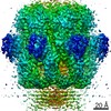
EMDB-19177:
Structure of the 55LCC ATPase complex

PDB-8rhn:
Structure of the 55LCC ATPase complex
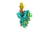
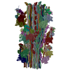
EMDB-29722:
Cryo-EM structure of the human cardiac myosin filament

EMDB-29726:
Cryo-EM structure of the human cardiac myosin filament

EMDB-29734:
Cryo-EM structure of the human cardiac myosin filament
PDB-8g4l:
Cryo-EM structure of the human cardiac myosin filament

EMDB-33752:
Cryo-EM structure of Mycobacterial Type VII Secretion System Virulence Factor EspB (residues 1-332) with Phosphatidic acid (PA)

EMDB-34878:
Cryo-EM structure of Mycobacterial Type VII Secretion System Virulence Factor EspB (residues 1-332)
EMDB-34391:
Octahedral supramolecular assembly of the bicomponent gamma-hemolysin octameric pore complexes from Staphylococcus aureus Newman.
PDB-8gz7:
Octahedral supramolecular assembly of the bicomponent gamma-hemolysin octameric pore complexes from Staphylococcus aureus Newman.

EMDB-33411:
Supramolecular assembly of Thermostable Direct Hemolysin from Vibrio parahaemolyticus

EMDB-33412:
Wild type Thermostable Direct Hemolysin (TDH) from Vibrio parahaemolyticus

EMDB-33413:
N-terminal deleted (NTD) Thermostable Direct Hemolysin from Vibrio parahaemolyticus

PDB-7yl9:
Cryo-EM structure of complete transmembrane channel E289A mutant Vibrio cholerae Cytolysin

EMDB-26993:
Cryo-EM structure of SARS-CoV-2 M protein in a lipid nanodisc

PDB-8ctk:
Cryo-EM structure of SARS-CoV-2 M protein in a lipid nanodisc
EMDB-33331:
Cyo-EM model for native cystathionine beta-synthase of Mycobacterium tuberculosis.
EMDB-33348:
Cryo-EM map of cystathionine beta-synthase of Mycobacterium tuberculosis in the presence of S-adenosylmethionine.
EMDB-33363:
Cryo-EM map of cystathionine beta-synthase of Mycobacterium tuberculosis in the presence of S-adenosylmethionine and serine.
PDB-7xnz:
Native cystathionine beta-synthase of Mycobacterium tuberculosis.
PDB-7xoh:
Cystathionine beta-synthase of Mycobacterium tuberculosis in the presence of S-adenosylmethionine.
PDB-7xoy:
Cystathionine beta-synthase of Mycobacterium tuberculosis in the presence of S-adenosylmethionine and serine.

EMDB-33215:
Cryo-EM reconstruction of complete transmembrane channel E289A mutant Vibrio cholerae Cytolysin

EMDB-33219:
Cryo-EM reconstruction of partial transmembrane channel E289A mutant Vibrio cholerae Cytolysin
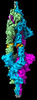
EMDB-32388:
Cryo-EM 3D model of the 3-RBD up dimeric spike protein of SARS-CoV2 in the presence of SIH-5
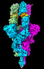
EMDB-33042:
Cryo-EM 3D model of the 3-RBD up single trimeric spike protein of SARS-CoV2 in the presence of synthetic peptide SIH-5.

PDB-7x7n:
3D model of the 3-RBD up single trimeric spike protein of SARS-CoV2 in the presence of synthetic peptide SIH-5.

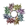
EMDB-32016:
Small heat shock protein (60-mer) from Synechococcus phage S-ShM2

EMDB-32017:
Small heat shock protein (48-mer) from Synechococcus phage S-ShM2

EMDB-31972:
Cryo-EM 3D reconstruction of Vibrio cholerae Cytolysin embedded in lipid bilayer- State 3
EMDB-31973:
Cryo-EM 3D reconstruction of Vibrio cholerae Cytolysin adhered to liposome membrane surface

EMDB-31974:
Cryo-EM 3D reconstruction of Vibrio cholerae Cytolysin partially embedded in lipid bilayer- State 2
EMDB-31092:
SARS-CoV2 Spike Protein structure at pH 6.5 with C1 Symmetry (Class 2)

EMDB-31093:
SARS-CoV2 Spike Protein structure at pH 6.5 with C1 Symmetry (Class 3)
EMDB-31094:
SARS-CoV2 Spike Protein structure at pH 6.5 with C1 Symmetry (Class 4)
EMDB-31095:
SARS-CoV2 Spike Protein structure at pH 6.5 with C1 Symmetry (Class 5)
EMDB-31096:
SARS-CoV2 Spike Protein structure at pH 7.4 with C1 Symmetry (Class 3)

EMDB-31097:
SARS-CoV2 Spike Protein structure at pH 7.4 with C1 Symmetry (Class 5)

EMDB-31098:
SARS-CoV2 Spike Protein structure at pH 7.4 with C3 Symmetry
EMDB-31099:
SARS-CoV2 Spike Protein structure at pH 8.0 with C1 Symmetry (Class 1)
EMDB-31100:
SARS-CoV2 Spike Protein structure at pH 7.4 with C1 Symmetry (Class 9)
EMDB-31101:
SARS-CoV2 Spike Protein structure at pH 7.4 with C1 Symmetry (Class 8)
EMDB-31102:
SARS-CoV2 Spike Protein structure at pH 8.0 with C1 Symmetry (Class 2)
ページ:
 wwPDBはEMDBデータモデルのバージョン3へ移行します
