Movie
Movie Controller


+ Open data

- Basic information
| Entry | Database: PDB / ID: 8g4l | |||||||||||||||
|---|---|---|---|---|---|---|---|---|---|---|---|---|---|---|---|---|
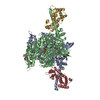
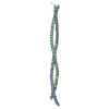
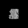
| Title | Cryo-EM structure of the human cardiac myosin filament | |||||||||||||||
 Components Components |
| |||||||||||||||
 Keywords Keywords | CONTRACTILE PROTEIN / cardiac / myosin / filament / complex | |||||||||||||||
| Function / homology |  Function and homology information Function and homology informationmyosin II heavy chain binding / C zone / regulation of muscle filament sliding / muscle cell fate specification / striated muscle myosin thick filament / regulation of slow-twitch skeletal muscle fiber contraction / sarcomerogenesis / titin-telethonin complex / regulation of the force of skeletal muscle contraction / structural molecule activity conferring elasticity ...myosin II heavy chain binding / C zone / regulation of muscle filament sliding / muscle cell fate specification / striated muscle myosin thick filament / regulation of slow-twitch skeletal muscle fiber contraction / sarcomerogenesis / titin-telethonin complex / regulation of the force of skeletal muscle contraction / structural molecule activity conferring elasticity / skeletal muscle myosin thick filament assembly / telethonin binding / regulation of striated muscle contraction / cardiac myofibril / detection of muscle stretch / muscle alpha-actinin binding / : / muscle myosin complex / A band / cardiac myofibril assembly / regulation of the force of heart contraction / transition between fast and slow fiber / myosin filament / cardiac muscle hypertrophy / cardiac muscle tissue morphogenesis / protein kinase regulator activity / mitotic chromosome condensation / Striated Muscle Contraction / muscle filament sliding / cardiac muscle hypertrophy in response to stress / regulation of cardiac muscle cell contraction / adult heart development / myosin complex / actinin binding / M band / myosin II complex / cardiac muscle cell development / I band / structural constituent of muscle / sarcomere organization / ventricular cardiac muscle tissue morphogenesis / myosin binding / microfilament motor activity / myosin heavy chain binding / heart contraction / positive regulation of the force of heart contraction / ATPase activator activity / myofibril / cytoskeletal motor activity / striated muscle thin filament / skeletal muscle thin filament assembly / actin monomer binding / skeletal muscle tissue development / ATP metabolic process / heart morphogenesis / striated muscle contraction / skeletal muscle contraction / cardiac muscle contraction / stress fiber / titin binding / regulation of heart rate / muscle contraction / condensed nuclear chromosome / post-embryonic development / positive regulation of protein secretion / sarcomere / negative regulation of cell growth / response to calcium ion / Z disc / actin filament binding / Platelet degranulation / actin cytoskeleton / heart development / actin binding / protein tyrosine kinase activity / protease binding / cytoskeleton / calmodulin binding / non-specific serine/threonine protein kinase / cell adhesion / protein serine kinase activity / protein serine/threonine kinase activity / calcium ion binding / positive regulation of gene expression / protein kinase binding / enzyme binding / protein homodimerization activity / extracellular exosome / extracellular region / ATP binding / metal ion binding / identical protein binding / cytoplasm / cytosol Similarity search - Function | |||||||||||||||
| Biological species |  Homo sapiens (human) Homo sapiens (human) | |||||||||||||||
| Method | ELECTRON MICROSCOPY / single particle reconstruction / cryo EM / Resolution: 6.4 Å | |||||||||||||||
 Authors Authors | Dutta, D. / Nguyen, V. / Padron, R. / Craig, R. | |||||||||||||||
| Funding support |  United States, 4items United States, 4items
| |||||||||||||||
 Citation Citation | Journal: Nature / Year: 2023 Title: Cryo-EM structure of the human cardiac myosin filament. Authors: Debabrata Dutta / Vu Nguyen / Kenneth S Campbell / Raúl Padrón / Roger Craig / Abstract: Pumping of the heart is powered by filaments of the motor protein myosin that pull on actin filaments to generate cardiac contraction. In addition to myosin, the filaments contain cardiac myosin- ...Pumping of the heart is powered by filaments of the motor protein myosin that pull on actin filaments to generate cardiac contraction. In addition to myosin, the filaments contain cardiac myosin-binding protein C (cMyBP-C), which modulates contractility in response to physiological stimuli, and titin, which functions as a scaffold for filament assembly. Myosin, cMyBP-C and titin are all subject to mutation, which can lead to heart failure. Despite the central importance of cardiac myosin filaments to life, their molecular structure has remained a mystery for 60 years. Here we solve the structure of the main (cMyBP-C-containing) region of the human cardiac filament using cryo-electron microscopy. The reconstruction reveals the architecture of titin and cMyBP-C and shows how myosin's motor domains (heads) form three different types of motif (providing functional flexibility), which interact with each other and with titin and cMyBP-C to dictate filament architecture and function. The packing of myosin tails in the filament backbone is also resolved. The structure suggests how cMyBP-C helps to generate the cardiac super-relaxed state; how titin and cMyBP-C may contribute to length-dependent activation; and how mutations in myosin and cMyBP-C might disturb interactions, causing disease. The reconstruction resolves past uncertainties and integrates previous data on cardiac muscle structure and function. It provides a new paradigm for interpreting structural, physiological and clinical observations, and for the design of potential therapeutic drugs. | |||||||||||||||
| History |
|
- Structure visualization
| Structure viewer | Molecule: MolmilJmol/JSmol |
|---|
- Downloads & links
-Download
| PDBx/mmCIF format | 8g4l.cif.gz | 21.3 MB | Display | PDBx/mmCIF format |
|---|---|---|---|---|
| PDB format | pdb8g4l.ent.gz | Display | PDB format | |
| PDBx/mmJSON format | 8g4l.json.gz | Tree view | PDBx/mmJSON format | |
| Others |  Other downloads Other downloads |
-Validation report
| Arichive directory | https://data.pdbj.org/pub/pdb/validation_reports/g4/8g4lftp://data.pdbj.org/pub/pdb/validation_reports/g4/8g4l | HTTPS FTP |
|---|
-Related structure data
| Related structure data | 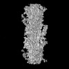 29722MC M: map data used to model this data C: citing same article ( |
|---|---|
| Similar structure data |

-Links
 PDBj
PDBj


- Assembly
| Deposited unit | 
|
|---|---|
| 1 |
|
-Components
| #1: Protein | Mass: 223445.984 Da / Num. of mol.: 78 / Source method: isolated from a natural source / Source: (natural) Homo sapiens (human) / Tissue: heart / References: UniProt: P12883#2: Protein | Mass: 21962.068 Da / Num. of mol.: 18 / Source method: isolated from a natural source / Source: (natural) Homo sapiens (human) / Tissue: heart / References: UniProt: P08590#3: Protein | Mass: 18813.273 Da / Num. of mol.: 18 / Source method: isolated from a natural source / Source: (natural) Homo sapiens (human) / Tissue: heart / References: UniProt: P10916#4: Protein | Mass: 119771.961 Da / Num. of mol.: 6 / Source method: isolated from a natural source / Source: (natural) Homo sapiens (human) / Tissue: heartReferences: UniProt: Q8WZ42, non-specific serine/threonine protein kinase #5: Protein | Mass: 140947.172 Da / Num. of mol.: 3 / Source method: isolated from a natural source / Source: (natural) Homo sapiens (human) / Tissue: heart / References: UniProt: Q14896Has protein modification | Y | |
|---|
-Experimental details
-Experiment
| Experiment | Method: ELECTRON MICROSCOPY |
|---|---|
| EM experiment | Aggregation state: FILAMENT / 3D reconstruction method: single particle reconstruction |
- Sample preparation
| Component | Name: Myosin filaments isolated from human cardiac left ventricular muscle Type: TISSUE / Entity ID: all / Source: NATURAL |
|---|---|
| Molecular weight | Value: 5.9 MDa / Experimental value: NO |
| Source (natural) | Organism: Homo sapiens (human) |
| Buffer solution | pH: 6.8 |
| Specimen | Embedding applied: NO / Shadowing applied: NO / Staining applied: NO / Vitrification applied: YES |
| Vitrification | Cryogen name: ETHANE |
- Electron microscopy imaging
| Experimental equipment |  Model: Titan Krios / Image courtesy: FEI Company |
|---|---|
| Microscopy | Model: FEI TITAN KRIOS |
| Electron gun | Electron source:  FIELD EMISSION GUN / Accelerating voltage: 300 kV / Illumination mode: FLOOD BEAM FIELD EMISSION GUN / Accelerating voltage: 300 kV / Illumination mode: FLOOD BEAM |
| Electron lens | Mode: BRIGHT FIELD / Nominal defocus max: 2000 nm / Nominal defocus min: 1000 nm |
| Image recording | Electron dose: 61 e/Å2 / Film or detector model: GATAN K3 (6k x 4k) |
- Processing
| Software |
| |||||||||||||||||||||||||||||||||||||||||||||||||||||||||||||||||||||||||||||||||||||||||||||||||||||||||||||||||||||||||||||||||||||||||||||||||||
|---|---|---|---|---|---|---|---|---|---|---|---|---|---|---|---|---|---|---|---|---|---|---|---|---|---|---|---|---|---|---|---|---|---|---|---|---|---|---|---|---|---|---|---|---|---|---|---|---|---|---|---|---|---|---|---|---|---|---|---|---|---|---|---|---|---|---|---|---|---|---|---|---|---|---|---|---|---|---|---|---|---|---|---|---|---|---|---|---|---|---|---|---|---|---|---|---|---|---|---|---|---|---|---|---|---|---|---|---|---|---|---|---|---|---|---|---|---|---|---|---|---|---|---|---|---|---|---|---|---|---|---|---|---|---|---|---|---|---|---|---|---|---|---|---|---|---|---|---|
| EM software |
| |||||||||||||||||||||||||||||||||||||||||||||||||||||||||||||||||||||||||||||||||||||||||||||||||||||||||||||||||||||||||||||||||||||||||||||||||||
| CTF correction | Type: PHASE FLIPPING AND AMPLITUDE CORRECTION | |||||||||||||||||||||||||||||||||||||||||||||||||||||||||||||||||||||||||||||||||||||||||||||||||||||||||||||||||||||||||||||||||||||||||||||||||||
| Symmetry | Point symmetry: C3 (3 fold cyclic) | |||||||||||||||||||||||||||||||||||||||||||||||||||||||||||||||||||||||||||||||||||||||||||||||||||||||||||||||||||||||||||||||||||||||||||||||||||
| 3D reconstruction | Resolution: 6.4 Å / Resolution method: FSC 0.143 CUT-OFF / Num. of particles: 102581 / Symmetry type: POINT | |||||||||||||||||||||||||||||||||||||||||||||||||||||||||||||||||||||||||||||||||||||||||||||||||||||||||||||||||||||||||||||||||||||||||||||||||||
| Atomic model building |
| |||||||||||||||||||||||||||||||||||||||||||||||||||||||||||||||||||||||||||||||||||||||||||||||||||||||||||||||||||||||||||||||||||||||||||||||||||
| Atomic model building |
|