 Movie
Movie Controller
 Structure viewers
 About EMN search


-Search query
-Search result
Showing 1 - 50 of 78 items for (author: cascio & p)

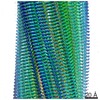
EMDB-70224: 
amyloid fibril of recombinant full-length 2N4R tau complexed with unfractionated mouse liver RNA and seeded by Alzheimer's disease tau fibrils
Method: helical / : Jiang YX, Sawaya MR, Abskharon R, Ge P, Boyer DR, Eisenberg DS

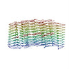
EMDB-70227:
amyloid fibril of recombinant full-length 2N4R tau complexed with mouse liver 18S ribosomal RNA
Method: helical / : Jiang YX, Sawaya MR, Abskharon R, Ge P, Boyer DR, Eisenberg DS

PDB-9o8e:
amyloid fibril of recombinant full-length 2N4R tau complexed with unfractionated mouse liver RNA and seeded by Alzheimer's disease tau fibrils
Method: helical / : Jiang YX, Sawaya MR, Abskharon R, Ge P, Boyer DR, Eisenberg DS

PDB-9o8h:
amyloid fibril of recombinant full-length 2N4R tau complexed with mouse liver 18S ribosomal RNA
Method: helical / : Jiang YX, Sawaya MR, Abskharon R, Ge P, Boyer DR, Eisenberg DS

EMDB-47731:
(Apo)Alpha-synuclein fibril structures from MSA patient brain
Method: helical / : Lu J, Sawaya MR, Ge P, Boyer DR, Eisenberg DS

EMDB-47732:
Complex fibril structure of MSA alpha-synuclein with CNS-11g at 4 hours
Method: helical / : Lu J, Sawaya MR, Ge P, Boyer DR, Eisenberg DS

EMDB-47733:
Complex fibril structure of MSA alpha-synuclein with CNS-11g at 6 hours
Method: helical / : Lu J, Sawaya MR, Ge P, Boyer DR, Eisenberg DS

EMDB-47734:
Complex fibril structure of MSA alpha-synuclein with CNS-11g at 10 hours
Method: helical / : Lu J, Sawaya MR, Ge P, Boyer DR, Eisenberg DS

EMDB-47735:
Complex fibril structure of MSA alpha-synuclein with CNS-11g at 15 hours
Method: helical / : Lu J, Sawaya MR, Ge P, Boyer DR, Eisenberg DS

EMDB-47736:
CNS-11g alone fibril
Method: helical / : Lu J, Sawaya MR
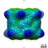
EMDB-15769:
A gap across the beta rings in 20S proteasome
Method: single particle / : Szenkier N, Arie M, Matzov D, Sertchook R, Carmeli R, Cascio P, Stanhill A, Shalev Benami M, Navon A
EMDB-15767:
Bovine 20S proteasome, untreated
Method: single particle / : Szenkier N, Arie M, Matzov D, Sertchook R, Carmeli R, Cascio P, Stanhill A, Shalev Benami M, Navon A
EMDB-15768:
Partially disassembled 20S proteasome upon disulfide bond formation.
Method: single particle / : Szenkier N, Arie M, Matzov D, Sertchook R, Carmeli R, Cascio P, Stanhill A, Shalev Benami M, Navon A

PDB-8azk:
Bovine 20S proteasome, untreated
Method: single particle / : Szenkier N, Arie M, Matzov D, Sertchook R, Carmeli R, Cascio P, Stanhill A, Shalev Benami M, Navon A
PDB-8t1n:
Micro-ED Structure of a Novel Domain of Unknown Function Solved with AlphaFold
Method: electron crystallography / : Miller JE, Cascio D, Sawaya MR, Cannon KA, Rodriguez JA, Yeates TO

EMDB-28014:
HnRNPA2 D290V LCD PM3
Method: helical / : Eisenberg DS, Lu J, Ge P, Boyer DR

PDB-8ec7:
HnRNPA2 D290V LCD PM3
Method: helical / : Eisenberg DS, Lu J, Ge P, Boyer DR

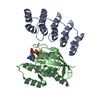
EMDB-29700:
Cryo-EM imaging scaffold subunits A and B used to display KRAS G12C complex with GDP
Method: single particle / : Castells-Graells R, Sawaya MR, Yeates TO
EMDB-29713:
KRAS G12C complex with GDP imaged on a cryo-EM imaging scaffold
Method: single particle / : Castells-Graells R, Sawaya MR, Yeates TO
EMDB-29715:
KRAS G12C complex with GDP and AMG 510 imaged on a cryo-EM imaging scaffold
Method: single particle / : Castells-Graells R, Sawaya MR, Yeates TO

EMDB-29718:
Green Fluorescence Protein imaged on a cryo-EM imaging scaffold
Method: single particle / : Castells-Graells R, Sawaya MR, Yeates TO
EMDB-29719:
KRAS G12V complex with GDP imaged on a cryo-EM imaging scaffold
Method: single particle / : Castells-Graells R, Sawaya MR, Yeates TO

EMDB-29720:
KRAS G13C complex with GDP imaged on a cryo-EM imaging scaffold
Method: single particle / : Castells-Graells R, Sawaya MR, Yeates TO
PDB-8g3k:
Cryo-EM imaging scaffold subunits A and B used to display KRAS G12C complex with GDP
Method: single particle / : Castells-Graells R, Sawaya MR, Yeates TO
PDB-8g42:
KRAS G12C complex with GDP imaged on a cryo-EM imaging scaffold
Method: single particle / : Castells-Graells R, Sawaya MR, Yeates TO
PDB-8g47:
KRAS G12C complex with GDP and AMG 510 imaged on a cryo-EM imaging scaffold
Method: single particle / : Castells-Graells R, Sawaya MR, Yeates TO
PDB-8g4e:
Green Fluorescence Protein imaged on a cryo-EM imaging scaffold
Method: single particle / : Castells-Graells R, Sawaya MR, Yeates TO
PDB-8g4f:
KRAS G12V complex with GDP imaged on a cryo-EM imaging scaffold
Method: single particle / : Castells-Graells R, Sawaya MR, Yeates TO
PDB-8g4h:
KRAS G13C complex with GDP imaged on a cryo-EM imaging scaffold
Method: single particle / : Castells-Graells R, Sawaya MR, Yeates TO

EMDB-27713:
HnRNPA2 D290V LCD PM1
Method: helical / : Eisenberg DS, Lu J, Ge P, Boyer DR

EMDB-27728:
HnRNPA2 D290V LCD PM2
Method: helical / : Eisenberg DS, Lu J, Ge P, Boyer DR

PDB-8du2:
HnRNPA2 D290V LCD PM1
Method: helical / : Eisenberg DS, Lu J, Ge P, Boyer DR

PDB-8duw:
HnRNPA2 D290V LCD PM2
Method: helical / : Eisenberg DS, Lu J, Ge P, Boyer DR

EMDB-21871:
hnRNPA2 Low complexity domain (LCD) determined by cryoEM
Method: helical / : Lu J, Cao Q

PDB-6wqk:
hnRNPA2 Low complexity domain (LCD) determined by cryoEM
Method: helical / : Lu J, Cao Q, Hughes MP, Sawaya MR, Boyer DR, Cascio D, Eisenberg DS

EMDB-0619:
Amyloid Beta KLVFFAENVGS 16-26 D23N Iowa mutation
Method: electron crystallography / : Griner SL, Sawaya MR

PDB-6o4j:
Amyloid Beta KLVFFAENVGS 16-26 D23N Iowa mutation
Method: electron crystallography / : Griner SL, Sawaya MR, Rodriguez JA, Cascio D, Gonen T
EMDB-9190:
Cell-free-expressed Pyridoxal 5'-phosphate synthase-like subunit (PDX1.2) from Arabidopsis thaliana
Method: single particle / : Novikova IV, Evans JE, Hellmann H, Sharma N

EMDB-7466:
Segment NFGTFS, with familial mutation A315T and phosphorylated threonine, from the low complexity domain of TDP-43, residues 312-317
Method: electron crystallography / : Guenther EL, Cao Q

EMDB-7467:
SWGMMGMLASQ segment from the low complexity domain of TDP-43, residues 333-343
Method: electron crystallography / : Guenther EL, Rodriguez JA

EMDB-8857:
MicroED structure of the segment, NFGEFS, from the A315E familial variant of the low complexity domain of TDP-43, residues 312-317
Method: electron crystallography / : Guenther EL, Sawaya MR

PDB-5wkb:
MicroED structure of the segment, NFGEFS, from the A315E familial variant of the low complexity domain of TDP-43, residues 312-317
Method: electron crystallography / : Guenther EL, Sawaya MR, Cascio D, Eisenberg DS

PDB-6cf4:
Segment NFGTFS, with familial mutation A315T and phosphorylated threonine, from the low complexity domain of TDP-43, residues 312-317
Method: electron crystallography / : Guenther EL, Cao Q, Boyer DR, Sawaya MR, Eisenberg DS

PDB-6cfh:
SWGMMGMLASQ segment from the low complexity domain of TDP-43
Method: electron crystallography / : Guenther EL, Rodriguez JA, Sawaya MR, Eisenberg DS

PDB-6bzm:
GFGNFGTS from low-complexity/FG repeat domain of Nup98, residues 116-123
Method: electron crystallography / : Hughes MP, Rodriguez JA, Sawaya MR, Cascio D, Chong L, Gonen T, Eisenberg DS

PDB-6bzp:
STGGYG from low-complexity domain of FUS, residues 77-82
Method: electron crystallography / : Hughes MP, Rodriguez JA, Sawaya MR, Cascio D, Gonen T, Eisenberg DS
EMDB-8781:
CryoEM structure of the segment, DLIIKGISVHI, assembled into a triple-helical fibril
Method: helical / : Guenther EL, Ge P
PDB-5w7v:
CryoEM structure of the segment, DLIIKGISVHI, assembled into a triple-helical fibril
Method: helical / : Guenther EL, Ge P, Eisenberg DS
EMDB-8765:
MicroED structure of the segment, DLIIKGISVHI, from the RRM2 of TDP-43, residues 247-257
Method: electron crystallography / : Guenther EL, Sawaya MR

PDB-5w52:
MicroED structure of the segment, DLIIKGISVHI, from the RRM2 of TDP-43, residues 247-257
Method: electron crystallography / : Guenther EL, Sawaya MR, Cascio D, Eisenberg DS
Pages:
 wwPDB to switch to version 3 of the EMDB data model
