 Movie
Movie Controller


+ Open data

- Basic information
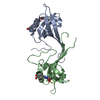
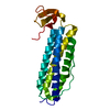
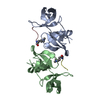
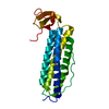
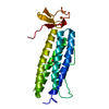
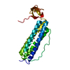
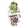
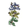
| Entry | Database: PDB / ID: 6wqk | |||||||||
|---|---|---|---|---|---|---|---|---|---|---|
| Title | hnRNPA2 Low complexity domain (LCD) determined by cryoEM | |||||||||
 Components Components | MCherry fluorescent protein,Heterogeneous nuclear ribonucleoproteins A2/B1 chimera | |||||||||
 Keywords Keywords | PROTEIN FIBRIL / Ribinucleoprotein / RNA binding protein / Low complexity domain | |||||||||
| Function / homology |  Function and homology information Function and homology informationmiRNA transport / positive regulation of telomere maintenance via telomere lengthening / RNA transport / DNA geometric change / primary miRNA processing / N6-methyladenosine-containing RNA reader activity / single-stranded telomeric DNA binding / G-rich strand telomeric DNA binding / miRNA binding / Processing of Capped Intron-Containing Pre-mRNA ...miRNA transport / positive regulation of telomere maintenance via telomere lengthening / RNA transport / DNA geometric change / primary miRNA processing / N6-methyladenosine-containing RNA reader activity / single-stranded telomeric DNA binding / G-rich strand telomeric DNA binding / miRNA binding / Processing of Capped Intron-Containing Pre-mRNA / mRNA transport / Cajal body / mRNA export from nucleus / catalytic step 2 spliceosome / DNA polymerase binding / mRNA Splicing - Major Pathway / Gene and protein expression by JAK-STAT signaling after Interleukin-12 stimulation / bioluminescence / spliceosomal complex / generation of precursor metabolites and energy / mRNA 3'-UTR binding / molecular condensate scaffold activity / mRNA splicing, via spliceosome / nuclear matrix / mRNA processing / chromosome, telomeric region / ribonucleoprotein complex / RNA binding / extracellular exosome / nucleoplasm / membrane / identical protein binding / nucleus / cytoplasm Similarity search - Function | |||||||||
| Biological species |  Anaplasma marginale (bacteria) Anaplasma marginale (bacteria) Homo sapiens (human) Homo sapiens (human) | |||||||||
| Method | ELECTRON MICROSCOPY / helical reconstruction / cryo EM / Resolution: 3.1 Å | |||||||||
 Authors Authors | Lu, J. / Cao, Q. / Hughes, M.P. / Sawaya, M.R. / Boyer, D.R. / Cascio, D. / Eisenberg, D.S. | |||||||||
| Funding support |  United States, 2items United States, 2items
| |||||||||
 Citation Citation | Journal: Nat Commun / Year: 2020 Title: CryoEM structure of the low-complexity domain of hnRNPA2 and its conversion to pathogenic amyloid. Authors: Jiahui Lu / Qin Cao / Michael P Hughes / Michael R Sawaya / David R Boyer / Duilio Cascio / David S Eisenberg / Abstract: hnRNPA2 is a human ribonucleoprotein (RNP) involved in RNA metabolism. It forms fibrils both under cellular stress and in mutated form in neurodegenerative conditions. Previous work established that ...hnRNPA2 is a human ribonucleoprotein (RNP) involved in RNA metabolism. It forms fibrils both under cellular stress and in mutated form in neurodegenerative conditions. Previous work established that the C-terminal low-complexity domain (LCD) of hnRNPA2 fibrillizes under stress, and missense mutations in this domain are found in the disease multisystem proteinopathy (MSP). However, little is known at the atomic level about the hnRNPA2 LCD structure that is involved in those processes and how disease mutations cause structural change. Here we present the cryo-electron microscopy (cryoEM) structure of the hnRNPA2 LCD fibril core and demonstrate its capability to form a reversible hydrogel in vitro containing amyloid-like fibrils. Whereas these fibrils, like pathogenic amyloid, are formed from protein chains stacked into β-sheets by backbone hydrogen bonds, they display distinct structural differences: the chains are kinked, enabling non-covalent cross-linking of fibrils and disfavoring formation of pathogenic steric zippers. Both reversibility and energetic calculations suggest these fibrils are less stable than pathogenic amyloid. Moreover, the crystal structure of the disease-mutation-containing segment (D290V) of hnRNPA2 suggests that the replacement fundamentally alters the fibril structure to a more stable energetic state. These findings illuminate how molecular interactions promote protein fibril networks and how mutation can transform fibril structure from functional to a pathogenic form. | |||||||||
| History |
|
- Structure visualization
| Movie |
Movie viewer |
|---|---|
| Structure viewer | Molecule: MolmilJmol/JSmol |
- Downloads & links
-Download
| PDBx/mmCIF format | 6wqk.cif.gz | 84.1 KB | Display | PDBx/mmCIF format |
|---|---|---|---|---|
| PDB format | pdb6wqk.ent.gz | 47.9 KB | Display | PDB format |
| PDBx/mmJSON format | 6wqk.json.gz | Tree view | PDBx/mmJSON format | |
| Others |  Other downloads Other downloads |
-Validation report
| Arichive directory | https://data.pdbj.org/pub/pdb/validation_reports/wq/6wqkftp://data.pdbj.org/pub/pdb/validation_reports/wq/6wqk | HTTPS FTP |
|---|
-Related structure data
| Related structure data |  21871MC  6wpqC C: citing same article ( M: map data used to model this data |
|---|---|
| Similar structure data | |
| EM raw data | EMPIAR-10634 (Title: CryoEM structure of the LCD of hnRNPA2 amyloid-like fibrils Data size: 1.2 TB Data #1: Unaligned micrographs, 1.15e-/A^2/frame [micrographs - multiframe]) |


-Links
 PDBj
PDBj


- Assembly
| Deposited unit | 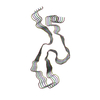
|
|---|---|
| 1 |
|
| Symmetry | Helical symmetry: (Circular symmetry: 1 / Dyad axis: no / N subunits divisor: 1 / Num. of operations: 5 / Rise per n subunits: 4.81 Å / Rotation per n subunits: -2.88 °) |
-Components
| #1: Protein | Mass: 45666.625 Da / Num. of mol.: 5 / Fragment: low complexity domain (UNP residues 193-353) Source method: isolated from a genetically manipulated source Source: (gene. exp.) Anaplasma marginale (bacteria), (gene. exp.) Homo sapiens (human)Gene: mCherry, HNRNPA2B1, HNRPA2B1 / Production host: |
|---|
-Experimental details
-Experiment
| Experiment | Method: ELECTRON MICROSCOPY |
|---|---|
| EM experiment | Aggregation state: FILAMENT / 3D reconstruction method: helical reconstruction |
- Sample preparation
| Component | Name: heterogeneous nuclear ribonucleo-protein A2 / Type: COMPLEX / Entity ID: all / Source: RECOMBINANT |
|---|---|
| Source (natural) | Organism: Homo sapiens (human) |
| Source (recombinant) | Organism: |
| Buffer solution | pH: 7.5 |
| Specimen | Embedding applied: NO / Shadowing applied: NO / Staining applied: NO / Vitrification applied: YES |
| Vitrification | Cryogen name: ETHANE |
- Electron microscopy imaging
| Experimental equipment |  Model: Titan Krios / Image courtesy: FEI Company |
|---|---|
| Microscopy | Model: FEI TITAN KRIOS |
| Electron gun | Electron source:  FIELD EMISSION GUN / Accelerating voltage: 300 kV / Illumination mode: OTHER FIELD EMISSION GUN / Accelerating voltage: 300 kV / Illumination mode: OTHER |
| Electron lens | Mode: BRIGHT FIELD |
| Image recording | Electron dose: 1.15 e/Å2 / Film or detector model: GATAN K2 IS (4k x 4k) |
- Processing
| Software | Name: PHENIX / Version: dev_3724: / Classification: refinement | ||||||||||||||||||||||||
|---|---|---|---|---|---|---|---|---|---|---|---|---|---|---|---|---|---|---|---|---|---|---|---|---|---|
| EM software |
| ||||||||||||||||||||||||
| CTF correction | Type: PHASE FLIPPING AND AMPLITUDE CORRECTION | ||||||||||||||||||||||||
| Helical symmerty | Angular rotation/subunit: -2.88 ° / Axial rise/subunit: 4.81 Å / Axial symmetry: C1 | ||||||||||||||||||||||||
| 3D reconstruction | Resolution: 3.1 Å / Resolution method: FSC 0.143 CUT-OFF / Num. of particles: 132571 / Symmetry type: HELICAL | ||||||||||||||||||||||||
| Refine LS restraints |
|
