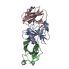
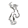
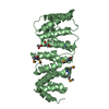
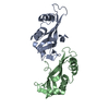
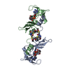
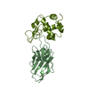
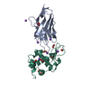
Entry Database : PDB / ID : 4criTitle Crystal Structure of 53BP1 tandem tudor domains in complex with methylated K810 Rb peptide RB1 PROTEIN TUMOR SUPPRESSOR P53-BINDING PROTEIN 1 Keywords / / Function / homology Function Domain/homology Component
/ / / / / / / / / / / / / / / / / / / / / / / / / / / / / / / / / / / / / / / / / / / / / / / / / / / / / / / / / / / / / / / / / / / / / / / / / / / / / / / / / / / / / / / / / / / / / / / / / / / / / / / / / / / / / / / / / / / / / / / / / / / / / / / / / / / / / / / / / / / / / / / / / / / Biological species HOMO SAPIENS (human)Method / / / Resolution : 2.35 Å Authors Krojer, T. / Johansson, C. / Gileadi, C. / Fedorov, O. / Carr, S. / La Thangue, N.B. / Vollmar, M. / Crawley, L. / von Delft, F. / Bountra, C. ...Krojer, T. / Johansson, C. / Gileadi, C. / Fedorov, O. / Carr, S. / La Thangue, N.B. / Vollmar, M. / Crawley, L. / von Delft, F. / Bountra, C. / Arrowsmith, C.H. / Edwards, A. / Oppermann, U. Journal : Proc.Natl.Acad.Sci.USA / Year : 2014Title : Lysine Methylation-Dependent Binding of 53BP1 to the Prb Tumor Suppressor.Authors : Carr, S.M. / Munro, S. / Zalmas, L. / Fedorov, O. / Johansson, C. / Krojer, T. / Sagum, C.A. / Bedford, M.T. / Oppermann, U. / La Thangue, N.B. History Deposition Feb 26, 2014 Deposition site / Processing site Revision 1.0 Aug 6, 2014 Provider / Type Revision 1.1 Aug 20, 2014 Group Revision 1.2 Dec 20, 2023 Group Data collection / Database references ... Data collection / Database references / Derived calculations / Other / Refinement description Category chem_comp_atom / chem_comp_bond ... chem_comp_atom / chem_comp_bond / database_2 / pdbx_database_status / pdbx_initial_refinement_model / struct_conn Item _database_2.pdbx_DOI / _database_2.pdbx_database_accession ... _database_2.pdbx_DOI / _database_2.pdbx_database_accession / _pdbx_database_status.status_code_sf / _struct_conn.pdbx_leaving_atom_flag
Show all Show less
 Movie
Movie Controller

 Yorodumi
 Open data
 Basic information
 Components
 Keywords
 Function and homology information
 HOMO SAPIENS (human)
 X-RAY DIFFRACTION /
  Authors
 Citation
 Structure visualization
 Downloads & links
 Other downloads
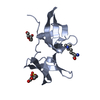


 PDBj
PDBj


 Assembly
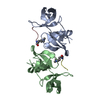

 Mass: 18.015 Da / Num. of mol.: 23 / Source method: isolated from a natural source / Formula: H2O
 Sample preparation
 / Beamline: I04 / Wavelength: 0.9795
  Processing