 Movie
Movie Controller


+ Open data

- Basic information
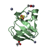
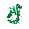
| Entry | Database: PDB / ID: 1i8n | ||||||
|---|---|---|---|---|---|---|---|
| Title | CRYSTAL STRUCTURE OF LEECH ANTI-PLATELET PROTEIN | ||||||
 Components Components | ANTI-PLATELET PROTEIN | ||||||
 Keywords Keywords | TOXIN / PAN MODULE | ||||||
| Function / homology | Hepatocyte Growth Factor / Hepatocyte Growth Factor / 3-Layer(bba) Sandwich / toxin activity / extracellular region / Alpha Beta / PROPIONAMIDE / Leech anti-platelet protein Function and homology information Function and homology information | ||||||
| Biological species |  Haementeria officinalis (Mexican leech) Haementeria officinalis (Mexican leech) | ||||||
| Method |  X-RAY DIFFRACTION / SYNCHROTRON / SIR / Resolution: 2.2 Å X-RAY DIFFRACTION / SYNCHROTRON / SIR / Resolution: 2.2 Å | ||||||
 Authors Authors | Huizinga, E.G. / Schouten, A. / Connolly, T.M. / Kroon, J. / Sixma, J.J. / Gros, P. | ||||||
 Citation Citation | Journal: Acta Crystallogr.,Sect.D / Year: 2001 Title: The structure of leech anti-platelet protein, an inhibitor of haemostasis. Authors: Huizinga, E.G. / Schouten, A. / Connolly, T.M. / Kroon, J. / Sixma, J.J. / Gros, P. #1: Journal: J.Biol.Chem. / Year: 1992Title: An inhibitor of Collagen-Stimulated Platelet Activation from the salivary glands of the Haementeria Officinalis Leech. I. Identification, Isolation, and Characterization Authors: Connolly, T.M. / Jacobs, J.W. / Condra, C. #2: Journal: J.Biol.Chem. / Year: 1992Title: An inhibitor of Collagen-Stimulated Platelet Activation from the salivary glands of the Haementeria Officinalis Leech. II. Cloning of the cDNA and Expression Authors: Keller, P.M. / Schultz, L.D. / Condra, C. / Karczewski, J. / Connolly, T.M. | ||||||
| History |
|
- Structure visualization
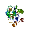
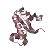
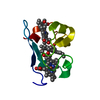
| Structure viewer | Molecule: MolmilJmol/JSmol |
|---|
- Downloads & links
-Download
| PDBx/mmCIF format | 1i8n.cif.gz | 71.2 KB | Display | PDBx/mmCIF format |
|---|---|---|---|---|
| PDB format | pdb1i8n.ent.gz | 52.7 KB | Display | PDB format |
| PDBx/mmJSON format | 1i8n.json.gz | Tree view | PDBx/mmJSON format | |
| Others |  Other downloads Other downloads |
-Validation report
| Arichive directory | https://data.pdbj.org/pub/pdb/validation_reports/i8/1i8nftp://data.pdbj.org/pub/pdb/validation_reports/i8/1i8n | HTTPS FTP |
|---|
-Related structure data
| Similar structure data |
|---|


-Links
 PDBj
PDBj- Assembly
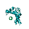
| Deposited unit | 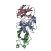
| ||||||||||||
|---|---|---|---|---|---|---|---|---|---|---|---|---|---|
| 1 | 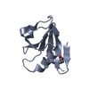
| ||||||||||||
| 2 | 
| ||||||||||||
| 3 | 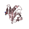
| ||||||||||||
| Unit cell |
| ||||||||||||
| Components on special symmetry positions |
|
-Components
| #1: Protein | Mass: 13767.517 Da / Num. of mol.: 3 Source method: isolated from a genetically manipulated source Source: (gene. exp.) Haementeria officinalis (Mexican leech)Gene: LAPP / Plasmid: PKH4 ALPHA 2 / Production host:  #2: Chemical | 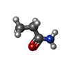  Mass: 73.094 Da / Num. of mol.: 3 / Source method: obtained synthetically / Formula: C3H7NO Mass: 73.094 Da / Num. of mol.: 3 / Source method: obtained synthetically / Formula: C3H7NO#3: Water | ChemComp-HOH / |  Mass: 18.015 Da / Num. of mol.: 190 / Source method: isolated from a natural source / Formula: H2O Mass: 18.015 Da / Num. of mol.: 190 / Source method: isolated from a natural source / Formula: H2OHas protein modification | Y | |
|---|
-Experimental details
-Experiment
| Experiment | Method: X-RAY DIFFRACTION / Number of used crystals: 1 |
|---|
- Sample preparation
| Crystal | Density Matthews: 3.36 Å3/Da / Density % sol: 63.4 % | |||||||||||||||||||||||||
|---|---|---|---|---|---|---|---|---|---|---|---|---|---|---|---|---|---|---|---|---|---|---|---|---|---|---|
| Crystal grow | Temperature: 277 K / Method: vapor diffusion, hanging drop / pH: 4.3 Details: MPD, sodium citrate, pH 4.3, VAPOR DIFFUSION, HANGING DROP, temperature 277K | |||||||||||||||||||||||||
| Crystal grow | *PLUS pH: 7 | |||||||||||||||||||||||||
| Components of the solutions | *PLUS
|
-Data collection
| Diffraction | Mean temperature: 100 K |
|---|---|
| Diffraction source | Source: SYNCHROTRON / Site: EMBL/DESY, HAMBURG  / Beamline: BW7B / Wavelength: 0.8825 Å / Beamline: BW7B / Wavelength: 0.8825 Å |
| Detector | Type: MARRESEARCH / Detector: IMAGE PLATE / Date: Jan 1, 1996 |
| Radiation | Monochromator: Si(111) / Protocol: SINGLE WAVELENGTH / Monochromatic (M) / Laue (L): M / Scattering type: x-ray |
| Radiation wavelength | Wavelength: 0.8825 Å / Relative weight: 1 |
| Reflection | Resolution: 2.2→20 Å / Num. all: 27864 / Num. obs: 27864 / % possible obs: 97.1 % / Observed criterion σ(F): 0 / Observed criterion σ(I): -3.7 / Redundancy: 4.4 % / Biso Wilson estimate: 36.4 Å2 / Rmerge(I) obs: 0.037 / Net I/σ(I): 35.6 |
| Reflection shell | Resolution: 2.2→2.25 Å / Redundancy: 3.8 % / Rmerge(I) obs: 0.253 / Mean I/σ(I) obs: 5.1 / Num. unique all: 1822 / Rsym value: 0.253 / % possible all: 96.5 |
| Reflection | *PLUS Lowest resolution: 20 Å |
| Reflection shell | *PLUS Lowest resolution: 2.23 Å / % possible obs: 96.5 % |
- Processing
| Software |
| ||||||||||||||||||||||||||||||||||||||||
|---|---|---|---|---|---|---|---|---|---|---|---|---|---|---|---|---|---|---|---|---|---|---|---|---|---|---|---|---|---|---|---|---|---|---|---|---|---|---|---|---|---|
| Refinement | Method to determine structure: SIR / Resolution: 2.2→20 Å / Isotropic thermal model: Isotropic / Cross valid method: THROUGHOUT / σ(F): 0 / σ(I): 0 / Stereochemistry target values: Engh & Huber
| ||||||||||||||||||||||||||||||||||||||||
| Solvent computation | Bsol: 42.9627 Å2 / ksol: 0.359435 e/Å3 | ||||||||||||||||||||||||||||||||||||||||
| Displacement parameters | Biso mean: 41.6 Å2
| ||||||||||||||||||||||||||||||||||||||||
| Refine analyze |
| ||||||||||||||||||||||||||||||||||||||||
| Refinement step | Cycle: LAST / Resolution: 2.2→20 Å
| ||||||||||||||||||||||||||||||||||||||||
| Refine LS restraints |
| ||||||||||||||||||||||||||||||||||||||||
| LS refinement shell | Resolution: 2.2→2.34 Å / Rfactor Rfree error: 0.018
| ||||||||||||||||||||||||||||||||||||||||
| Software | *PLUS Name: CNS / Version: 1 / Classification: refinement | ||||||||||||||||||||||||||||||||||||||||
| Refinement | *PLUS Highest resolution: 2.2 Å / Lowest resolution: 20 Å / σ(F): 0 / Rfactor obs: 0.215 / Rfactor Rfree: 0.24 | ||||||||||||||||||||||||||||||||||||||||
| Solvent computation | *PLUS | ||||||||||||||||||||||||||||||||||||||||
| Displacement parameters | *PLUS Biso mean: 41.6 Å2 | ||||||||||||||||||||||||||||||||||||||||
| Refine LS restraints | *PLUS
| ||||||||||||||||||||||||||||||||||||||||
| LS refinement shell | *PLUS Rfactor Rfree: 0.332 / Rfactor Rwork: 0.312 |
