 Movie
Movie Controller


+ Open data

- Basic information
| Entry | Database: PDB / ID: 5ixl | ||||||
|---|---|---|---|---|---|---|---|
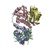
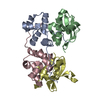
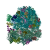
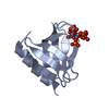
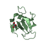
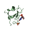
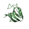
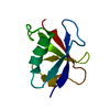
| Title | Structure of P. vulgaris HigB toxin Y91A variant | ||||||
 Components Components | Endoribonuclease HigB | ||||||
 Keywords Keywords | HYDROLASE / bacterial toxins / biofilms / cell metabolism / energy metabolism / microbial pathogenesis / stress response / stringent response / translation control | ||||||
| Function / homology |  Function and homology information Function and homology informationplasmid maintenance / RNA catabolic process / RNA endonuclease activity / translation repressor activity / negative regulation of cell growth / ribosome binding / Hydrolases; Acting on ester bonds / negative regulation of translation / negative regulation of cell population proliferation / hydrolase activity / mRNA binding Similarity search - Function | ||||||
| Biological species |  Proteus vulgaris (bacteria) Proteus vulgaris (bacteria) | ||||||
| Method |  X-RAY DIFFRACTION / SYNCHROTRON / MOLECULAR REPLACEMENT / Resolution: 1.55 Å X-RAY DIFFRACTION / SYNCHROTRON / MOLECULAR REPLACEMENT / Resolution: 1.55 Å | ||||||
 Authors Authors | Schureck, M.A. / Repack, A.A. / Miles, S.J. / Marquez, J. / Dunham, C.M. | ||||||
 Citation Citation | Journal: Nucleic Acids Res. / Year: 2016 Title: Mechanism of endonuclease cleavage by the HigB toxin. Authors: Schureck, M.A. / Repack, A. / Miles, S.J. / Marquez, J. / Dunham, C.M. | ||||||
| History |
|
- Structure visualization
| Structure viewer | Molecule: MolmilJmol/JSmol |
|---|
- Downloads & links
-Download
| PDBx/mmCIF format | 5ixl.cif.gz | 330.6 KB | Display | PDBx/mmCIF format |
|---|---|---|---|---|
| PDB format | pdb5ixl.ent.gz | 267.9 KB | Display | PDB format |
| PDBx/mmJSON format | 5ixl.json.gz | Tree view | PDBx/mmJSON format | |
| Others |  Other downloads Other downloads |
-Validation report
| Arichive directory | https://data.pdbj.org/pub/pdb/validation_reports/ix/5ixlftp://data.pdbj.org/pub/pdb/validation_reports/ix/5ixl | HTTPS FTP |
|---|
-Related structure data
| Related structure data |  4zsnC  5iwhC 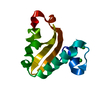 4px8S S: Starting model for refinement C: citing same article ( |
|---|---|
| Similar structure data |


-Links
 PDBj
PDBj- Assembly
| Deposited unit | 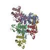
| ||||||||
|---|---|---|---|---|---|---|---|---|---|
| 1 | 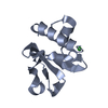
| ||||||||
| 2 | 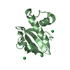
| ||||||||
| 3 | 
| ||||||||
| 4 | 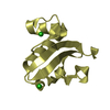
| ||||||||
| 5 | 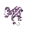
| ||||||||
| 6 | 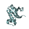
| ||||||||
| 7 | 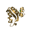
| ||||||||
| 8 | 
| ||||||||
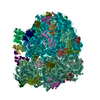
| Unit cell |
|
-Components
| #1: Protein | Mass: 13621.312 Da / Num. of mol.: 8 Source method: isolated from a genetically manipulated source Source: (gene. exp.) Proteus vulgaris (bacteria) / Gene: higB / Plasmid: pBAD/Myc-HisA / Production host: References: UniProt: Q7A225, Hydrolases; Acting on ester bonds #2: Chemical | ChemComp-CL /   Mass: 35.453 Da / Num. of mol.: 12 / Source method: obtained synthetically / Formula: Cl Mass: 35.453 Da / Num. of mol.: 12 / Source method: obtained synthetically / Formula: Cl#3: Water | ChemComp-HOH / |  Mass: 18.015 Da / Num. of mol.: 608 / Source method: isolated from a natural source / Formula: H2O Mass: 18.015 Da / Num. of mol.: 608 / Source method: isolated from a natural source / Formula: H2O |
|---|
-Experimental details
-Experiment
| Experiment | Method: X-RAY DIFFRACTION / Number of used crystals: 1 |
|---|
- Sample preparation
| Crystal | Density Matthews: 1.55 Å3/Da / Density % sol: 20.68 % |
|---|---|
| Crystal grow | Temperature: 293.15 K / Method: vapor diffusion, sitting drop / pH: 7.5 Details: 40% (w/v) polyethylene glycol monomethylether 2,000 and 0.15 M KBr |
-Data collection
| Diffraction | Mean temperature: 100 K |
|---|---|
| Diffraction source | Source: SYNCHROTRON / Site: APS  / Beamline: 22-ID / Wavelength: 1 Å / Beamline: 22-ID / Wavelength: 1 Å |
| Detector | Type: RAYONIX MX300-HS / Detector: CCD / Date: Feb 15, 2015 |
| Radiation | Monochromator: Cryogenically-cooled Si-220 / Protocol: SINGLE WAVELENGTH / Monochromatic (M) / Laue (L): M / Scattering type: x-ray |
| Radiation wavelength | Wavelength: 1 Å / Relative weight: 1 |
| Reflection | Resolution: 1.55→100 Å / Num. obs: 87109 / % possible obs: 85.7 % / Redundancy: 2.6 % / Net I/σ(I): 12 |
| Reflection shell | Resolution: 1.55→1.6 Å / Redundancy: 2.5 % / Mean I/σ(I) obs: 2.4 / % possible all: 89.9 |
- Processing
| Software |
| |||||||||||||||||||||||||||||||||||||||||||||||||||||||||||||||||||||||||||||||||||||||||||
|---|---|---|---|---|---|---|---|---|---|---|---|---|---|---|---|---|---|---|---|---|---|---|---|---|---|---|---|---|---|---|---|---|---|---|---|---|---|---|---|---|---|---|---|---|---|---|---|---|---|---|---|---|---|---|---|---|---|---|---|---|---|---|---|---|---|---|---|---|---|---|---|---|---|---|---|---|---|---|---|---|---|---|---|---|---|---|---|---|---|---|---|---|
| Refinement | Method to determine structure: MOLECULAR REPLACEMENT Starting model: 4PX8 Resolution: 1.55→37.004 Å / SU ML: 0.18 / Cross valid method: FREE R-VALUE / σ(F): 1.35 / Phase error: 22.55
| |||||||||||||||||||||||||||||||||||||||||||||||||||||||||||||||||||||||||||||||||||||||||||
| Solvent computation | Shrinkage radii: 0.9 Å / VDW probe radii: 1.11 Å | |||||||||||||||||||||||||||||||||||||||||||||||||||||||||||||||||||||||||||||||||||||||||||
| Refinement step | Cycle: LAST / Resolution: 1.55→37.004 Å
| |||||||||||||||||||||||||||||||||||||||||||||||||||||||||||||||||||||||||||||||||||||||||||
| Refine LS restraints |
| |||||||||||||||||||||||||||||||||||||||||||||||||||||||||||||||||||||||||||||||||||||||||||
| LS refinement shell |
|
