 Movie
Movie Controller


+ Open data

- Basic information
| Entry | Database: EMDB / ID: EMD-21871 | |||||||||
|---|---|---|---|---|---|---|---|---|---|---|
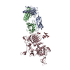
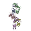
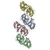
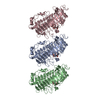
| Title | hnRNPA2 Low complexity domain (LCD) determined by cryoEM | |||||||||
 Map data Map data | ||||||||||
 Sample Sample |
| |||||||||
 Keywords Keywords | Ribinucleoprotein / RNA binding protein / Low complexity domain / PROTEIN FIBRIL | |||||||||
| Function / homology |  Function and homology information Function and homology informationmiRNA transport / positive regulation of telomere maintenance via telomere lengthening / RNA transport / DNA geometric change / primary miRNA processing / N6-methyladenosine-containing RNA reader activity / single-stranded telomeric DNA binding / G-rich strand telomeric DNA binding / miRNA binding / Processing of Capped Intron-Containing Pre-mRNA ...miRNA transport / positive regulation of telomere maintenance via telomere lengthening / RNA transport / DNA geometric change / primary miRNA processing / N6-methyladenosine-containing RNA reader activity / single-stranded telomeric DNA binding / G-rich strand telomeric DNA binding / miRNA binding / Processing of Capped Intron-Containing Pre-mRNA / mRNA transport / Cajal body / mRNA export from nucleus / catalytic step 2 spliceosome / DNA polymerase binding / mRNA Splicing - Major Pathway / Gene and protein expression by JAK-STAT signaling after Interleukin-12 stimulation / bioluminescence / spliceosomal complex / generation of precursor metabolites and energy / mRNA 3'-UTR binding / molecular condensate scaffold activity / mRNA splicing, via spliceosome / nuclear matrix / mRNA processing / chromosome, telomeric region / ribonucleoprotein complex / RNA binding / extracellular exosome / nucleoplasm / membrane / identical protein binding / nucleus / cytoplasm Similarity search - Function | |||||||||
| Biological species |  Homo sapiens (human) Homo sapiens (human) | |||||||||
| Method | helical reconstruction / cryo EM / Resolution: 3.1 Å | |||||||||
 Authors Authors | Lu J / Cao Q | |||||||||
| Funding support |  United States, 2 items United States, 2 items
| |||||||||
 Citation Citation | Journal: Nat Commun / Year: 2020 Title: CryoEM structure of the low-complexity domain of hnRNPA2 and its conversion to pathogenic amyloid. Authors: Jiahui Lu / Qin Cao / Michael P Hughes / Michael R Sawaya / David R Boyer / Duilio Cascio / David S Eisenberg / Abstract: hnRNPA2 is a human ribonucleoprotein (RNP) involved in RNA metabolism. It forms fibrils both under cellular stress and in mutated form in neurodegenerative conditions. Previous work established that ...hnRNPA2 is a human ribonucleoprotein (RNP) involved in RNA metabolism. It forms fibrils both under cellular stress and in mutated form in neurodegenerative conditions. Previous work established that the C-terminal low-complexity domain (LCD) of hnRNPA2 fibrillizes under stress, and missense mutations in this domain are found in the disease multisystem proteinopathy (MSP). However, little is known at the atomic level about the hnRNPA2 LCD structure that is involved in those processes and how disease mutations cause structural change. Here we present the cryo-electron microscopy (cryoEM) structure of the hnRNPA2 LCD fibril core and demonstrate its capability to form a reversible hydrogel in vitro containing amyloid-like fibrils. Whereas these fibrils, like pathogenic amyloid, are formed from protein chains stacked into β-sheets by backbone hydrogen bonds, they display distinct structural differences: the chains are kinked, enabling non-covalent cross-linking of fibrils and disfavoring formation of pathogenic steric zippers. Both reversibility and energetic calculations suggest these fibrils are less stable than pathogenic amyloid. Moreover, the crystal structure of the disease-mutation-containing segment (D290V) of hnRNPA2 suggests that the replacement fundamentally alters the fibril structure to a more stable energetic state. These findings illuminate how molecular interactions promote protein fibril networks and how mutation can transform fibril structure from functional to a pathogenic form. | |||||||||
| History |
|
- Structure visualization
| Movie |
Movie viewer |
|---|---|
| Structure viewer | EM map: SurfViewMolmilJmol/JSmol |
| Supplemental images |

- Downloads & links
-EMDB archive
| Map data | emd_21871.map.gz | 7.4 MB | EMDB map data format | |
|---|---|---|---|---|
| Header (meta data) | emd-21871-v30.xmlemd-21871.xml | 9.7 KB 9.7 KB | Display Display | EMDB header |
| Images | 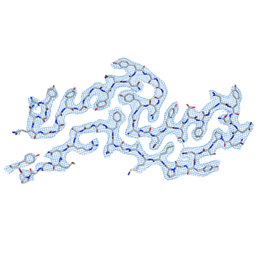 emd_21871.png emd_21871.png | 141.8 KB | ||
| Filedesc metadata | emd-21871.cif.gz | 5.4 KB | ||
| Archive directory |  http://ftp.pdbj.org/pub/emdb/structures/EMD-21871ftp://ftp.pdbj.org/pub/emdb/structures/EMD-21871 http://ftp.pdbj.org/pub/emdb/structures/EMD-21871ftp://ftp.pdbj.org/pub/emdb/structures/EMD-21871 | HTTPS FTP |
-Related structure data
| Related structure data |  6wqkMC  6wpqC M: atomic model generated by this map C: citing same article ( |
|---|---|
| Similar structure data | |
| EM raw data | EMPIAR-10634 (Title: CryoEM structure of the LCD of hnRNPA2 amyloid-like fibrils Data size: 1.2 TB Data #1: Unaligned micrographs, 1.15e-/A^2/frame [micrographs - multiframe]) |


-Links
| EMDB pages | EMDB (EBI/PDBe) / EMDataResource |
|---|---|
| Related items in Molecule of the Month |


-Map
| File | Download / File: emd_21871.map.gz / Format: CCP4 / Size: 8 MB / Type: IMAGE STORED AS FLOATING POINT NUMBER (4 BYTES) | ||||||||||||||||||||||||||||||||||||||||||||||||||||||||||||||||||||
|---|---|---|---|---|---|---|---|---|---|---|---|---|---|---|---|---|---|---|---|---|---|---|---|---|---|---|---|---|---|---|---|---|---|---|---|---|---|---|---|---|---|---|---|---|---|---|---|---|---|---|---|---|---|---|---|---|---|---|---|---|---|---|---|---|---|---|---|---|---|
| Projections & slices | Image control
Images are generated by Spider. | ||||||||||||||||||||||||||||||||||||||||||||||||||||||||||||||||||||
| Voxel size | X=Y=Z: 1.064 Å | ||||||||||||||||||||||||||||||||||||||||||||||||||||||||||||||||||||
| Density |
| ||||||||||||||||||||||||||||||||||||||||||||||||||||||||||||||||||||
| Symmetry | Space group: 1 | ||||||||||||||||||||||||||||||||||||||||||||||||||||||||||||||||||||
| Details | EMDB XML:
CCP4 map header:
| ||||||||||||||||||||||||||||||||||||||||||||||||||||||||||||||||||||
 X (Sec.)
 Y (Row.)
 Z (Col.)


-Supplemental data
- Sample components
-Entire : heterogeneous nuclear ribonucleo-protein A2
| Entire | Name: heterogeneous nuclear ribonucleo-protein A2 |
|---|---|
| Components |
|
-Supramolecule #1: heterogeneous nuclear ribonucleo-protein A2
| Supramolecule | Name: heterogeneous nuclear ribonucleo-protein A2 / type: complex / ID: 1 / Parent: 0 / Macromolecule list: all |
|---|---|
| Source (natural) | Organism: Homo sapiens (human) |
-Macromolecule #1: MCherry fluorescent protein,Heterogeneous nuclear ribonucleoprote...
| Macromolecule | Name: MCherry fluorescent protein,Heterogeneous nuclear ribonucleoproteins A2/B1 chimera type: protein_or_peptide / ID: 1 / Number of copies: 5 / Enantiomer: LEVO |
|---|---|
| Source (natural) | Organism: Homo sapiens (human) |
| Molecular weight | Theoretical: 45.666625 KDa |
| Recombinant expression | Organism:  |
| Sequence | String: MSYYHHHHHH DYDIPTTENL YFQGAMVSKG EEDNMAIIKE FMRFKVHMEG SVNGHEFEIE GEGEGRPYEG TQTAKLKVTK GGPLPFAWD ILSPQFMYGS KAYVKHPADI PDYLKLSFPE GFKWERVMNF EDGGVVTVTQ DSSLQDGEFI YKVKLRGTNF P SDGPVMQK ...String: MSYYHHHHHH DYDIPTTENL YFQGAMVSKG EEDNMAIIKE FMRFKVHMEG SVNGHEFEIE GEGEGRPYEG TQTAKLKVTK GGPLPFAWD ILSPQFMYGS KAYVKHPADI PDYLKLSFPE GFKWERVMNF EDGGVVTVTQ DSSLQDGEFI YKVKLRGTNF P SDGPVMQK KTMGWEASSE RMYPEDGALK GEIKQRLKLK DGGHYDAEVK TTYKAKKPVQ LPGAYNVNIK LDITSHNEDY TI VEQYERA EGRHSTGGMD ELYKAMDPMQ EVQSSRSGRG GNFGFGDSRG GGGNFGPGPG SNFRGGSDGY GSGRGFGDGY NGY GGGPGG GNFGGSPGYG GGRGGYGGGG PGYGNQGGGY GGGYDNYGGG NYGSGNYNDF GNYNQQPSNY GPMKSGNFGG SRNM GGPYG GGNYGPGGSG GSGGYGGRSR Y UniProtKB: MCherry fluorescent protein, Heterogeneous nuclear ribonucleoproteins A2/B1 |
-Experimental details
-Structure determination
| Method | cryo EM |
|---|---|
 Processing Processing | helical reconstruction |
| Aggregation state | filament |
-Sample preparation
| Buffer | pH: 7.5 |
|---|---|
| Vitrification | Cryogen name: ETHANE |
- Electron microscopy
| Microscope | FEI TITAN KRIOS |
|---|---|
| Image recording | Film or detector model: GATAN K2 IS (4k x 4k) / Average electron dose: 1.15 e/Å2 |
| Electron beam | Acceleration voltage: 300 kV / Electron source:  FIELD EMISSION GUN FIELD EMISSION GUN |
| Electron optics | Illumination mode: OTHER / Imaging mode: BRIGHT FIELD |
| Experimental equipment |  Model: Titan Krios / Image courtesy: FEI Company |
-Image processing
| Final reconstruction | Applied symmetry - Helical parameters - Δz: 4.81 Å Applied symmetry - Helical parameters - Δ&Phi: -2.88 ° Applied symmetry - Helical parameters - Axial symmetry: C1 (asymmetric) Resolution.type: BY AUTHOR / Resolution: 3.1 Å / Resolution method: FSC 0.143 CUT-OFF / Number images used: 132571 |
|---|---|
| Startup model | Type of model: OTHER / Details: an elongated Gaussian blob |
| Final angle assignment | Type: NOT APPLICABLE |
