 Movie
Movie Controller

[English] 日本語
 Yorodumi
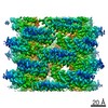
- EMDB-9190: Cell-free-expressed Pyridoxal 5'-phosphate synthase-like subunit ... -
+ Open data

- Basic information
| Entry | Database: EMDB / ID: EMD-9190 | |||||||||
|---|---|---|---|---|---|---|---|---|---|---|
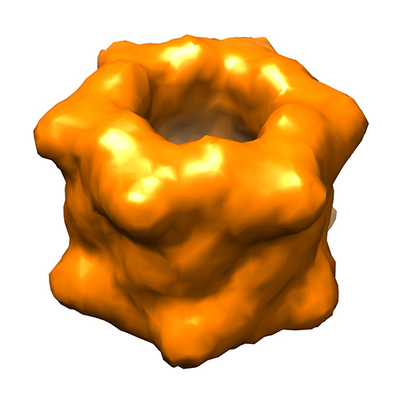
| Title | Cell-free-expressed Pyridoxal 5'-phosphate synthase-like subunit (PDX1.2) from Arabidopsis thaliana | |||||||||
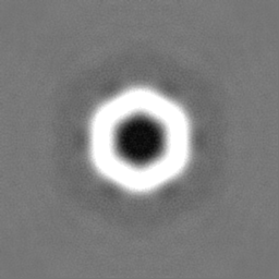
 Map data Map data | Cell-free-expressed PDX1.2 single particle cryo-EM map | |||||||||
 Sample Sample |
| |||||||||
| Biological species |  | |||||||||
| Method | single particle reconstruction / cryo EM / Resolution: 15.0 Å | |||||||||
 Authors Authors | Novikova IV / Evans JE / Hellmann H / Sharma N | |||||||||
 Citation Citation | Journal: Adv Struct Chem Imaging / Year: 2018 Title: Protein structural biology using cell-free platform from wheat germ. Authors: Irina V Novikova / Noopur Sharma / Trevor Moser / Ryan Sontag / Yan Liu / Michael J Collazo / Duilio Cascio / Tolou Shokuhfar / Hanjo Hellmann / Michael Knoblauch / James E Evans /  Abstract: One of the biggest bottlenecks for structural analysis of proteins remains the creation of high-yield and high-purity samples of the target protein. Cell-free protein synthesis technologies are ...One of the biggest bottlenecks for structural analysis of proteins remains the creation of high-yield and high-purity samples of the target protein. Cell-free protein synthesis technologies are powerful and customizable platforms for obtaining functional proteins of interest in short timeframes, while avoiding potential toxicity issues and permitting high-throughput screening. These methods have benefited many areas of genomic and proteomics research, therapeutics, vaccine development and protein chip constructions. In this work, we demonstrate a versatile and multiscale eukaryotic wheat germ cell-free protein expression pipeline to generate functional proteins of different sizes from multiple host organism and DNA source origins. We also report on a robust purification procedure, which can produce highly pure (> 98%) proteins with no specialized equipment required and minimal time invested. This pipeline successfully produced and analyzed proteins in all three major geometry formats used for structural biology including single particle analysis with electron microscopy, and both two-dimensional and three-dimensional protein crystallography. The flexibility of the wheat germ system in combination with the multiscale pipeline described here provides a new workflow for rapid production and purification of samples that may not be amenable to other recombinant approaches for structural characterization. | |||||||||
| History |
|
- Structure visualization
| Movie |
 Movie viewer Movie viewer |
|---|---|
| Structure viewer | EM map: SurfViewMolmilJmol/JSmol |
| Supplemental images |
- Downloads & links
-EMDB archive
| Map data | emd_9190.map.gz | 59.3 MB | EMDB map data format | |
|---|---|---|---|---|
| Header (meta data) | emd-9190-v30.xmlemd-9190.xml | 15.5 KB 15.5 KB | Display Display | EMDB header |
| FSC (resolution estimation) | emd_9190_fsc.xml | 10.7 KB | Display | FSC data file |
| Images |  emd_9190.png emd_9190.png | 154.9 KB | ||
| Others | emd_9190_half_map_1.map.gzemd_9190_half_map_2.map.gz | 5.4 MB 5.4 MB | ||
| Archive directory |  http://ftp.pdbj.org/pub/emdb/structures/EMD-9190ftp://ftp.pdbj.org/pub/emdb/structures/EMD-9190 http://ftp.pdbj.org/pub/emdb/structures/EMD-9190ftp://ftp.pdbj.org/pub/emdb/structures/EMD-9190 | HTTPS FTP |
-Related structure data
| Similar structure data |
|---|

-Links
| EMDB pages | EMDB (EBI/PDBe) / EMDataResource |
|---|
-Map
| File | Download / File: emd_9190.map.gz / Format: CCP4 / Size: 64 MB / Type: IMAGE STORED AS FLOATING POINT NUMBER (4 BYTES) | ||||||||||||||||||||||||||||||||||||||||||||||||||||||||||||
|---|---|---|---|---|---|---|---|---|---|---|---|---|---|---|---|---|---|---|---|---|---|---|---|---|---|---|---|---|---|---|---|---|---|---|---|---|---|---|---|---|---|---|---|---|---|---|---|---|---|---|---|---|---|---|---|---|---|---|---|---|---|
| Annotation | Cell-free-expressed PDX1.2 single particle cryo-EM map | ||||||||||||||||||||||||||||||||||||||||||||||||||||||||||||
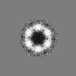
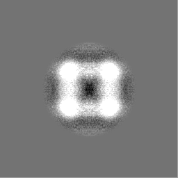
| Projections & slices | Image control
Images are generated by Spider. | ||||||||||||||||||||||||||||||||||||||||||||||||||||||||||||
| Voxel size | X=Y=Z: 1.172 Å | ||||||||||||||||||||||||||||||||||||||||||||||||||||||||||||
| Density |
| ||||||||||||||||||||||||||||||||||||||||||||||||||||||||||||
| Symmetry | Space group: 1 | ||||||||||||||||||||||||||||||||||||||||||||||||||||||||||||
| Details | EMDB XML:
CCP4 map header:
| ||||||||||||||||||||||||||||||||||||||||||||||||||||||||||||
 Z (Sec.)
 Y (Row.)
 X (Col.)


-Supplemental data
-Half map: Cell-free-expressed PDX1.2 single particle cryo-EM even half-map
| File | emd_9190_half_map_1.map | ||||||||||||
|---|---|---|---|---|---|---|---|---|---|---|---|---|---|
| Annotation | Cell-free-expressed PDX1.2 single particle cryo-EM even half-map | ||||||||||||
| Projections & Slices |
| ||||||||||||
| Density Histograms |


-Half map: Cell-free-expressed PDX1.2 single particle cryo-EM odd half-map
| File | emd_9190_half_map_2.map | ||||||||||||
|---|---|---|---|---|---|---|---|---|---|---|---|---|---|
| Annotation | Cell-free-expressed PDX1.2 single particle cryo-EM odd half-map | ||||||||||||
| Projections & Slices |
| ||||||||||||
| Density Histograms |


- Sample components
-Entire : Pyridoxal 5'-phosphate synthase-like subunit PDX1.2 with N-termin...
| Entire | Name: Pyridoxal 5'-phosphate synthase-like subunit PDX1.2 with N-terminal 3XFLAG tag |
|---|---|
| Components |
|
-Supramolecule #1: Pyridoxal 5'-phosphate synthase-like subunit PDX1.2 with N-termin...
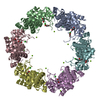
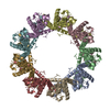
| Supramolecule | Name: Pyridoxal 5'-phosphate synthase-like subunit PDX1.2 with N-terminal 3XFLAG tag type: complex / ID: 1 / Parent: 0 / Macromolecule list: all / Details: Complex assembled from 12 monomers in D6 symmetry |
|---|---|
| Source (natural) | Organism: |
| Recombinant expression | Organism: |
| Molecular weight | Theoretical: 440 KDa |
-Macromolecule #1: Pyridoxal 5'-phosphate synthase-like subunit PDX1.2 with N-termin...
| Macromolecule | Name: Pyridoxal 5'-phosphate synthase-like subunit PDX1.2 with N-terminal 3XFLAG tag type: protein_or_peptide / ID: 1 / Enantiomer: DEXTRO EC number: pyridoxal 5'-phosphate synthase (glutamine hydrolysing) |
|---|---|
| Source (natural) | Organism: |
| Recombinant expression | Organism: |
| Sequence | String: MDYKDHDGDY KDHDIDYKDD DDKLAADQAM TDQDQGAVTL YSGTAITDAK KNHPFSVKVG LAQVLRGGA IVEVSSVNQA KLAESAGACS VIVSDPVRSR GGVRRMPDPV LIKEVKRAVS V PVMARARV GHFVEAQILE SLAVDYIDES EIISVADDDH FINKHNFRSP ...String: MDYKDHDGDY KDHDIDYKDD DDKLAADQAM TDQDQGAVTL YSGTAITDAK KNHPFSVKVG LAQVLRGGA IVEVSSVNQA KLAESAGACS VIVSDPVRSR GGVRRMPDPV LIKEVKRAVS V PVMARARV GHFVEAQILE SLAVDYIDES EIISVADDDH FINKHNFRSP FICGCRDTGE AL RRIREGA AMIRIQGDLT ATGNIAETVK NVRSLMGEVR VLNNMDDDEV FTFAKKISAP YDL VAQTKQ MGRVPVVQFA SGGITTPADA ALMMQLGCDG VFVGSEVFDG PDPFKKLRSI VQAV QHYND PHVLAEMSSG LENAMESLNV RGDRIQDFGQ GSV |
-Experimental details
-Structure determination
| Method | cryo EM |
|---|---|
 Processing Processing | single particle reconstruction |
| Aggregation state | particle |
-Sample preparation
| Concentration | 0.05 mg/mL | |||||||||
|---|---|---|---|---|---|---|---|---|---|---|
| Buffer | pH: 7.5 Component:
| |||||||||
| Grid | Model: C-flat-1.2/1.3 / Material: COPPER / Support film - Material: CARBON / Support film - topology: HOLEY / Pretreatment - Type: GLOW DISCHARGE / Pretreatment - Atmosphere: AIR / Pretreatment - Pressure: 0.026 kPa Details: Glow discharging conditions on EasyGlow: 10 mA, 30 sec, 0.26 mbar | |||||||||
| Vitrification | Cryogen name: ETHANE / Chamber humidity: 80 % / Chamber temperature: 298 K / Instrument: LEICA EM GP / Details: deposition: 3 uL sample, 3 sec blot. |
- Electron microscopy
| Microscope | JEOL 3000SFF |
|---|---|
| Image recording | Film or detector model: DIRECT ELECTRON DE-20 (5k x 3k) / Detector mode: INTEGRATING / Number real images: 42 / Average electron dose: 2.0 e/Å2 |
| Electron beam | Acceleration voltage: 300 kV / Electron source:  FIELD EMISSION GUN FIELD EMISSION GUN |
| Electron optics | Illumination mode: FLOOD BEAM / Imaging mode: BRIGHT FIELD / Cs: 1.6 mm |

