 ムービー
ムービー コントローラー
 構造ビューア
 EMN検索について


-検索条件
-検索結果
検索 (著者・登録者: baran & k)の結果全48件を表示しています
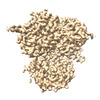
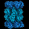
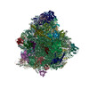
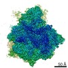
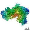
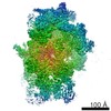
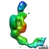
EMDB-41363: 
Cryo-EM structure of DDB1deltaB-DDA1-DCAF5

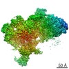
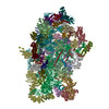
PDB-8tl6:
Cryo-EM structure of DDB1deltaB-DDA1-DCAF5

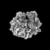
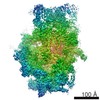
EMDB-17391:
Subtomogram average of ribosomes in germinated polar tubes of Vairimorpha necatrix

EMDB-17467:
Subtomogram representing a segment of the outer wall of empty germinated polar tubes from Vairimorpha necatrix.
EMDB-17468:
Subtomogram representing a segment of the outer wall of cargo-filled, germinated polar tubes from Vairimorpha necatrix.
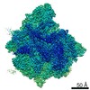
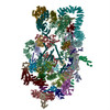
EMDB-15311:
Dedicated chaperone at the ribosome safeguards the proteostasis network during eEF1A biogenesis

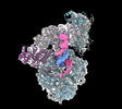
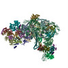
EMDB-27256:
Human DNA polymerase alpha/primase elongation complex I bound to primer/template
EMDB-27258:
Human DNA polymerase-alpha/primase elongation complex II bound to primer/template
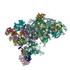
PDB-8d96:
Human DNA polymerase alpha/primase elongation complex I bound to primer/template
PDB-8d9d:
Human DNA polymerase-alpha/primase elongation complex II bound to primer/template
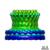
EMDB-14630:
Membrane-bound CHMP2A-CHMP3 filament (430 Angstrom diameter)
EMDB-14631:
Membrane-bound CHMP2A-CHMP3 filament (410 Angstrom diameter)
PDB-7zcg:
CHMP2A-CHMP3 heterodimer (430 Angstrom diameter)
PDB-7zch:
CHMP2A-CHMP3 heterodimer (410 Angstrom diameter)
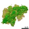
EMDB-15365:
Vairimorpha necatrix 20S proteasome from spores
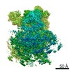
EMDB-15366:
Vairimorpha necatrix 26S proteasome from sporoplasms

EMDB-15367:
Vairimorpha necatrix 20S proteasome from sporoplasms
PDB-8adn:
Vairimorpha necatrix 20S proteasome from spores

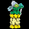
EMDB-12816:
Enterococcus faecalis EfrCD in complex with a nanobody

PDB-7ocy:
Enterococcus faecalis EfrCD in complex with a nanobody
EMDB-13776:
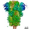
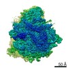
Structure of formaldehyde cross-linked SARS-CoV-2 S glycoprotein
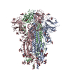
PDB-7q1z:
Structure of formaldehyde cross-linked SARS-CoV-2 S glycoprotein

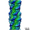
EMDB-13185:
Helical structure of the toxin MakA from Vibrio cholera

PDB-7p3r:
Helical structure of the toxin MakA from Vibrio cholera
EMDB-11437:
Structure of the Paranosema locustae ribosome in complex with Lso2
PDB-6zu5:
Structure of the Paranosema locustae ribosome in complex with Lso2
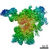
EMDB-4931:
Cryo-EM structure of the microsporidian ribosome: Multibody-refined map body 1 (LSU)
EMDB-4932:
Cryo-EM structure of the microsporidian ribosome: Multibody-refined map body 2 (SSU-body)

EMDB-4933:
Cryo-EM structure of the microsporidian ribosome: Multibody-refined map body 3 (SSU-head)
EMDB-4934:
Cryo-EM structure of the microsporidian ribosome: State 1, Multibody-refined map body 3 (SSU-head)
EMDB-4935:
Evolutionary compaction and adaptation visualized by the structure of the dormant microsporidian ribosome
PDB-6rm3:
Evolutionary compaction and adaptation visualized by the structure of the dormant microsporidian ribosome
EMDB-0441:
Conformational switches control early maturation of the eukaryotic small ribosomal subunit
PDB-6nd4:
Conformational switches control early maturation of the eukaryotic small ribosomal subunit
EMDB-7324:
Yeast nucleolar pre-60S ribosomal subunit (state 2)
EMDB-7445:
Yeast nucleolar pre-60S ribosomal subunit (state 3)
PDB-6c0f:
Yeast nucleolar pre-60S ribosomal subunit (state 2)
PDB-6cb1:
Yeast nucleolar pre-60S ribosomal subunit (state 3)
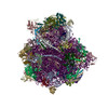
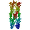
EMDB-8859:
The complete structure of the small subunit processome
PDB-5wlc:
The complete structure of the small subunit processome
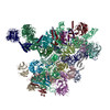
EMDB-8473:
Architecture of the yeast small subunit processome
PDB-5tzs:
Architecture of the yeast small subunit processome
EMDB-8223:
Random conical tilt reconstruction of Saccharomyces cerevisiae UtpB
EMDB-6370:
3D-Structure of negatively stained Schistosome myosin filament obtained by low-dose electron microscopy
PDB-3jax:
Heavy meromyosin from Schistosoma mansoni muscle thick filament by negative stain EM
EMDB-1769:
Perforin Pore

EMDB-1772:
Perforin monomer, conformation 1

EMDB-1773:
Perforin monomer, conformation 2
 wwPDBはEMDBデータモデルのバージョン3へ移行します
