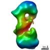
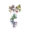
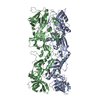
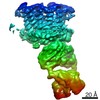
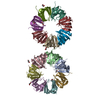
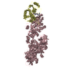
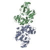
Journal: Nature / Year: 2010 Title: The structural basis for membrane binding and pore formation by lymphocyte perforin. Authors: Ruby H P Law / Natalya Lukoyanova / Ilia Voskoboinik / Tom T Caradoc-Davies / Katherine Baran / Michelle A Dunstone / Michael E D'Angelo / Elena V Orlova / Fasséli Coulibaly / Sandra ...Authors: Ruby H P Law / Natalya Lukoyanova / Ilia Voskoboinik / Tom T Caradoc-Davies / Katherine Baran / Michelle A Dunstone / Michael E D'Angelo / Elena V Orlova / Fasséli Coulibaly / Sandra Verschoor / Kylie A Browne / Annette Ciccone / Michael J Kuiper / Phillip I Bird / Joseph A Trapani / Helen R Saibil / James C Whisstock / Abstract: Natural killer cells and cytotoxic T lymphocytes accomplish the critically important function of killing virus-infected and neoplastic cells. They do this by releasing the pore-forming protein ...Natural killer cells and cytotoxic T lymphocytes accomplish the critically important function of killing virus-infected and neoplastic cells. They do this by releasing the pore-forming protein perforin and granzyme proteases from cytoplasmic granules into the cleft formed between the abutting killer and target cell membranes. Perforin, a 67-kilodalton multidomain protein, oligomerizes to form pores that deliver the pro-apoptopic granzymes into the cytosol of the target cell. The importance of perforin is highlighted by the fatal consequences of congenital perforin deficiency, with more than 50 different perforin mutations linked to familial haemophagocytic lymphohistiocytosis (type 2 FHL). Here we elucidate the mechanism of perforin pore formation by determining the X-ray crystal structure of monomeric murine perforin, together with a cryo-electron microscopy reconstruction of the entire perforin pore. Perforin is a thin 'key-shaped' molecule, comprising an amino-terminal membrane attack complex perforin-like (MACPF)/cholesterol dependent cytolysin (CDC) domain followed by an epidermal growth factor (EGF) domain that, together with the extreme carboxy-terminal sequence, forms a central shelf-like structure. A C-terminal C2 domain mediates initial, Ca(2+)-dependent membrane binding. Most unexpectedly, however, electron microscopy reveals that the orientation of the perforin MACPF domain in the pore is inside-out relative to the subunit arrangement in CDCs. These data reveal remarkable flexibility in the mechanism of action of the conserved MACPF/CDC fold and provide new insights into how related immune defence molecules such as complement proteins assemble into pores.
History
Deposition
Aug 3, 2010
-
Header (metadata) release
Sep 24, 2010
-
Map release
Nov 3, 2010
-
Update
Nov 16, 2010
-
Current status
Nov 16, 2010
Processing site: PDBe / Status: Released
-
Structure visualization
Movie
Surface view with section colored by density value
Type: NEGATIVE Details: 1% w/v uranyl acetate for 10 sec at room temperature
Grid
Details: Home made continuous carbon on 400 mesh copper grid
Vitrification
Cryogen name: NONE / Instrument: OTHER
-
Electron microscopy
Microscope
FEI TECNAI 12
Alignment procedure
Legacy - Astigmatism: At the specimen level
Date
May 18, 2007
Image recording
Category: FILM / Film or detector model: KODAK SO-163 FILM / Digitization - Scanner: ZEISS SCAI / Digitization - Sampling interval: 7 µm / Number real images: 32 / Average electron dose: 20 e/Å2 / Bits/pixel: 8
Electron beam
Acceleration voltage: 120 kV / Electron source: LAB6
Specimen holder: Side entry single tilt / Specimen holder model: SIDE ENTRY, EUCENTRIC
-
Image processing
Details
Particles were selected manually using EMAN-Boxer software
CTF correction
Details: Estimated with CTFFIND3, then phases flipped for each particle
Final reconstruction
Applied symmetry - Point group: C1 (asymmetric) / Algorithm: OTHER / Resolution.type: BY AUTHOR / Resolution: 25.0 Å / Resolution method: FSC 0.5 CUT-OFF / Software - Name: Imagic, Spider / Details: The hand of the map has not been determined / Number images used: 3400
In the structure databanks used in Yorodumi, some data are registered as the other names, "COVID-19 virus" and "2019-nCoV". Here are the details of the virus and the list of structure data.
Jan 31, 2019. EMDB accession codes are about to change! (news from PDBe EMDB page)
EMDB accession codes are about to change! (news from PDBe EMDB page)
The allocation of 4 digits for EMDB accession codes will soon come to an end. Whilst these codes will remain in use, new EMDB accession codes will include an additional digit and will expand incrementally as the available range of codes is exhausted. The current 4-digit format prefixed with “EMD-” (i.e. EMD-XXXX) will advance to a 5-digit format (i.e. EMD-XXXXX), and so on. It is currently estimated that the 4-digit codes will be depleted around Spring 2019, at which point the 5-digit format will come into force.
The EM Navigator/Yorodumi systems omit the EMD- prefix.
Related info.:Q: What is EMD? / ID/Accession-code notation in Yorodumi/EM Navigator
Yorodumi is a browser for structure data from EMDB, PDB, SASBDB, etc.
This page is also the successor to EM Navigator detail page, and also detail information page/front-end page for Omokage search.
The word "yorodu" (or yorozu) is an old Japanese word meaning "ten thousand". "mi" (miru) is to see.
Related info.:EMDB / PDB / SASBDB / Comparison of 3 databanks / Yorodumi Search / Aug 31, 2016. New EM Navigator & Yorodumi / Yorodumi Papers / Jmol/JSmol / Function and homology information / Changes in new EM Navigator and Yorodumi
 Movie
Movie Controller


 Open data
 Basic information
 Map data
 Sample
 Keywords
 Function and homology information
 Authors
 Citation
 Structure visualization
 Downloads & links
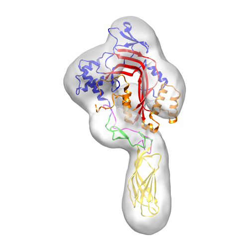 1772image.png
 http://ftp.pdbj.org/pub/emdb/structures/EMD-1772
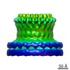

 Z (Sec.)
 Y (Row.)
 X (Col.)


 Sample components
 Spodoptera frugiperda (fall armyworm)
 Processing
 Electron microscopy
 UCSF Chimera