 Movie
Movie Controller

[English] 日本語
 Yorodumi
- PDB-1jri: The Crystal Structure of an Sm-like Archaeal Protein with Two Hep... -
+ Open data

- Basic information
| Entry | Database: PDB / ID: 1jri | ||||||
|---|---|---|---|---|---|---|---|
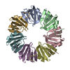
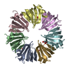
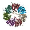
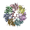
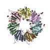
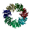
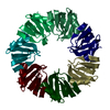
| Title | The Crystal Structure of an Sm-like Archaeal Protein with Two Heptamers in the Asymmetric Unit. | ||||||
 Components Components | Sm-like Archaeal Protein 1 (SmAP1) | ||||||
 Keywords Keywords | STRUCTURAL GENOMICS / UNKNOWN FUNCTION / heptameric / 35-stranded beta toroid | ||||||
| Function / homology |  Function and homology information Function and homology informationmRNA splicing, via spliceosome / ribonucleoprotein complex / RNA binding Similarity search - Function | ||||||
| Biological species |   Methanothermobacter thermautotrophicus (archaea) Methanothermobacter thermautotrophicus (archaea) | ||||||
| Method |  X-RAY DIFFRACTION / MOLECULAR REPLACEMENT / Resolution: 2.8 Å X-RAY DIFFRACTION / MOLECULAR REPLACEMENT / Resolution: 2.8 Å | ||||||
 Authors Authors | Mura, C. / Eisenberg, D. | ||||||
 Citation Citation | Journal: Protein Sci. / Year: 2003 Title: The oligomerization and ligand-binding properties of Sm-like archaeal proteins (SmAPs) Authors: Mura, C. / Kozhukhovsky, A. / Gingery, M. / Phillips, M. / Eisenberg, D. | ||||||
| History |
|
- Structure visualization
| Structure viewer | Molecule: MolmilJmol/JSmol |
|---|
- Downloads & links
-Download
| PDBx/mmCIF format | 1jri.cif.gz | 208.5 KB | Display | PDBx/mmCIF format |
|---|---|---|---|---|
| PDB format | pdb1jri.ent.gz | 171.1 KB | Display | PDB format |
| PDBx/mmJSON format | 1jri.json.gz | Tree view | PDBx/mmJSON format | |
| Others |  Other downloads Other downloads |
-Validation report
| Arichive directory | https://data.pdbj.org/pub/pdb/validation_reports/jr/1jriftp://data.pdbj.org/pub/pdb/validation_reports/jr/1jri | HTTPS FTP |
|---|
-Related structure data
| Related structure data |  1jbmSC 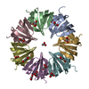 1lnxC 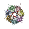 1lojC S: Starting model for refinement C: citing same article ( |
|---|---|
| Similar structure data |


-Links
 PDBj
PDBj

- Assembly
| Deposited unit | 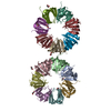
| ||||||||
|---|---|---|---|---|---|---|---|---|---|
| 1 | 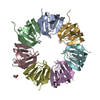
| ||||||||
| 2 | 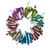
| ||||||||
| Unit cell |
| ||||||||
| Details | The asymmetric unit of the P212121 lattice contains two copies of the biologically significant assembly (a heptamer), the tangential rings being roughly coplanar (~15 degrees deviation). |
-Components
| #1: Protein | Mass: 9502.921 Da / Num. of mol.: 14 Source method: isolated from a genetically manipulated source Source: (gene. exp.) Methanothermobacter thermautotrophicus (archaea)Gene: Mth0649 / Species (production host): Escherichia coli / Production host:  #2: Chemical |   Mass: 35.453 Da / Num. of mol.: 3 / Source method: obtained synthetically / Formula: Cl Mass: 35.453 Da / Num. of mol.: 3 / Source method: obtained synthetically / Formula: Cl#3: Chemical | ChemComp-EDO /   Mass: 62.068 Da / Num. of mol.: 13 / Source method: obtained synthetically / Formula: C2H6O2 Mass: 62.068 Da / Num. of mol.: 13 / Source method: obtained synthetically / Formula: C2H6O2#4: Water | ChemComp-HOH / |  Mass: 18.015 Da / Num. of mol.: 86 / Source method: isolated from a natural source / Formula: H2O Mass: 18.015 Da / Num. of mol.: 86 / Source method: isolated from a natural source / Formula: H2O |
|---|
-Experimental details
-Experiment
| Experiment | Method: X-RAY DIFFRACTION / Number of used crystals: 1 |
|---|
- Sample preparation
| Crystal | Density Matthews: 2.08 Å3/Da / Density % sol: 40.74 % | |||||||||||||||||||||||||||||||||||||||||||||||||
|---|---|---|---|---|---|---|---|---|---|---|---|---|---|---|---|---|---|---|---|---|---|---|---|---|---|---|---|---|---|---|---|---|---|---|---|---|---|---|---|---|---|---|---|---|---|---|---|---|---|---|
| Crystal grow | Temperature: 292.8 K / Method: vapor diffusion, hanging drop / pH: 8.5 Details: 0.1 M Tris, 10% v/v isopropanol, pH 8.5, VAPOR DIFFUSION, HANGING DROP, temperature 19.8K | |||||||||||||||||||||||||||||||||||||||||||||||||
| Crystal grow | *PLUS pH: 7.8 | |||||||||||||||||||||||||||||||||||||||||||||||||
| Components of the solutions | *PLUS
|
-Data collection
| Diffraction | Mean temperature: 110 K |
|---|---|
| Diffraction source | Source: ROTATING ANODE / Type: RIGAKU / Wavelength: 1.5418 Å |
| Detector | Type: ADSC QUANTUM 4 / Detector: CCD / Date: Apr 1, 2001 / Details: fine-focussing mirrors |
| Radiation | Monochromator: Ni filter / Protocol: SINGLE WAVELENGTH / Monochromatic (M) / Laue (L): M / Scattering type: x-ray |
| Radiation wavelength | Wavelength: 1.5418 Å / Relative weight: 1 |
| Reflection | Resolution: 2.8→100 Å / Num. obs: 28487 / % possible obs: 99 % / Observed criterion σ(F): 0 / Observed criterion σ(I): -3 / Redundancy: 11.5 % / Rmerge(I) obs: 0.112 / Net I/σ(I): 19.3 |
| Reflection shell | Resolution: 2.8→2.9 Å / Rmerge(I) obs: 0.387 / Mean I/σ(I) obs: 3.3 / Num. unique all: 2645 / % possible all: 92.5 |
| Reflection | *PLUS Highest resolution: 2.8 Å / Num. measured all: 329838 |
| Reflection shell | *PLUS % possible obs: 92.5 % |
- Processing
| Software |
| |||||||||||||||||||||||||
|---|---|---|---|---|---|---|---|---|---|---|---|---|---|---|---|---|---|---|---|---|---|---|---|---|---|---|
| Refinement | Method to determine structure: MOLECULAR REPLACEMENT Starting model: PDB ENTRY 1JBM Resolution: 2.8→14.95 Å Isotropic thermal model: restrained isotropic temperature factors Cross valid method: THROUGHOUT / σ(F): 0 / σ(I): 0 / Stereochemistry target values: Engh & Huber
| |||||||||||||||||||||||||
| Displacement parameters | Biso mean: 52.3 Å2 | |||||||||||||||||||||||||
| Refine analyze |
| |||||||||||||||||||||||||
| Refinement step | Cycle: LAST / Resolution: 2.8→14.95 Å
| |||||||||||||||||||||||||
| Refine LS restraints |
| |||||||||||||||||||||||||
| LS refinement shell | Resolution: 2.8→2.97 Å / Rfactor Rfree error: 0.026
| |||||||||||||||||||||||||
| Software | *PLUS Name: CNS / Version: 1 / Classification: refinement | |||||||||||||||||||||||||
| Refinement | *PLUS σ(F): 0 / % reflection Rfree: 5 % / Rfactor obs: 0.199 / Rfactor Rfree: 0.29 | |||||||||||||||||||||||||
| Solvent computation | *PLUS | |||||||||||||||||||||||||
| Displacement parameters | *PLUS Biso mean: 52.3 Å2 | |||||||||||||||||||||||||
| Refine LS restraints | *PLUS
| |||||||||||||||||||||||||
| LS refinement shell | *PLUS Highest resolution: 2.8 Å / Rfactor Rfree: 0.375 / Rfactor Rwork: 0.28 |
