 Movie
Movie Controller

[English] 日本語
 Yorodumi
- EMDB-17467: Subtomogram representing a segment of the outer wall of empty ger... -
+ Open data

- Basic information
| Entry |  | |||||||||
|---|---|---|---|---|---|---|---|---|---|---|
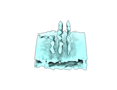
| Title | Subtomogram representing a segment of the outer wall of empty germinated polar tubes from Vairimorpha necatrix. | |||||||||
 Map data Map data | Merged map after refinement. | |||||||||
 Sample Sample |
| |||||||||
 Keywords Keywords | Polar tube / Subtomogram / Microsporidia / CELL INVASION | |||||||||
| Biological species |  Vairimorpha necatrix (fungus) Vairimorpha necatrix (fungus) | |||||||||
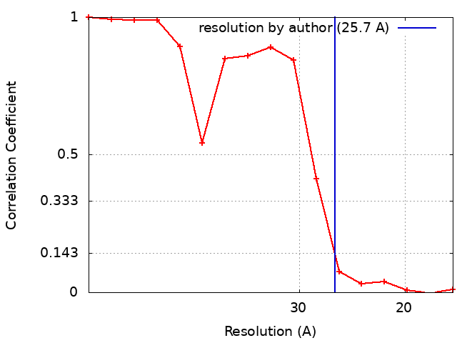
| Method | subtomogram averaging / cryo EM / Resolution: 25.7 Å | |||||||||
 Authors Authors | Sharma H / Ehrenbolger K / Jespersen N / Carlson LA / Barandun J | |||||||||
| Funding support | European Union, 2 items
| |||||||||
 Citation Citation | Journal: Biorxiv / Year: 2023 Title: Ribosome clustering and surface layer reorganization in the microsporidian host-invasion apparatus Authors: Sharma H / Jespersen N / Ehrenbolger K / Carlson LA / Barandun J | |||||||||
| History |
|
- Structure visualization
| Supplemental images |
|---|
- Downloads & links
-EMDB archive
| Map data | emd_17467.map.gz | 118.4 KB |  EMDB map data format EMDB map data format | |
|---|---|---|---|---|
| Header (meta data) | emd-17467-v30.xmlemd-17467.xml | 15.8 KB 15.8 KB | Display Display | EMDB header |
| FSC (resolution estimation) | emd_17467_fsc.xml | 1.6 KB | Display | FSC data file |
| Images |  emd_17467.png emd_17467.png | 39.8 KB | ||
| Masks | emd_17467_msk_1.map | 129 KB | Mask map | |
| Filedesc metadata | emd-17467.cif.gz | 4.4 KB | ||
| Others | emd_17467_half_map_1.map.gzemd_17467_half_map_2.map.gz | 118.7 KB 118.7 KB | ||
| Archive directory |  http://ftp.pdbj.org/pub/emdb/structures/EMD-17467ftp://ftp.pdbj.org/pub/emdb/structures/EMD-17467 http://ftp.pdbj.org/pub/emdb/structures/EMD-17467ftp://ftp.pdbj.org/pub/emdb/structures/EMD-17467 | HTTPS FTP |
-Related structure data


-Links
| EMDB pages | EMDB (EBI/PDBe) / EMDataResource |
|---|
-Map
| File | Download / File: emd_17467.map.gz / Format: CCP4 / Size: 128.9 KB / Type: IMAGE STORED AS FLOATING POINT NUMBER (4 BYTES) | ||||||||||||||||||||||||||||||||||||
|---|---|---|---|---|---|---|---|---|---|---|---|---|---|---|---|---|---|---|---|---|---|---|---|---|---|---|---|---|---|---|---|---|---|---|---|---|---|
| Annotation | Merged map after refinement. | ||||||||||||||||||||||||||||||||||||
| Projections & slices | Image control
Images are generated by Spider. | ||||||||||||||||||||||||||||||||||||
| Voxel size | X=Y=Z: 8.69 Å | ||||||||||||||||||||||||||||||||||||
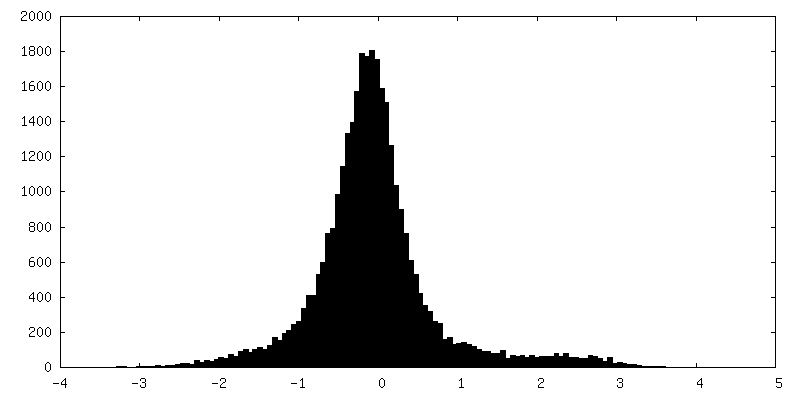
| Density |
| ||||||||||||||||||||||||||||||||||||
| Symmetry | Space group: 1 | ||||||||||||||||||||||||||||||||||||
| Details | EMDB XML:
|
 Z (Sec.)
 Y (Row.)
 X (Col.)


-Supplemental data
-Mask #1
| File | emd_17467_msk_1.map | ||||||||||||
|---|---|---|---|---|---|---|---|---|---|---|---|---|---|
| Projections & Slices |
| ||||||||||||
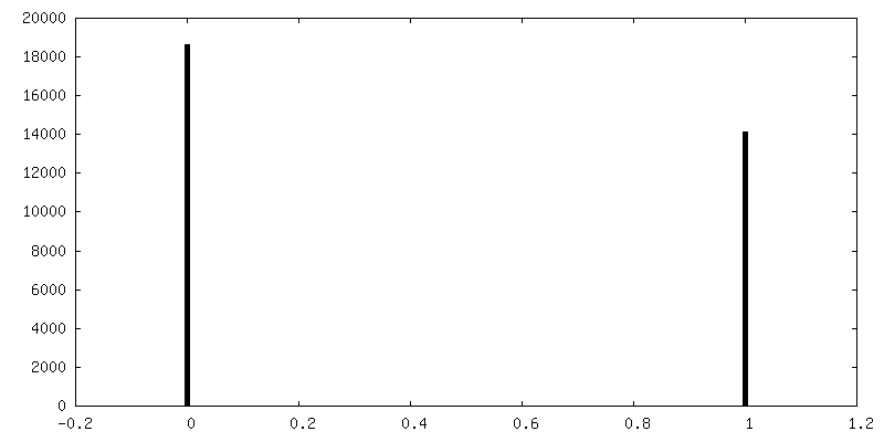
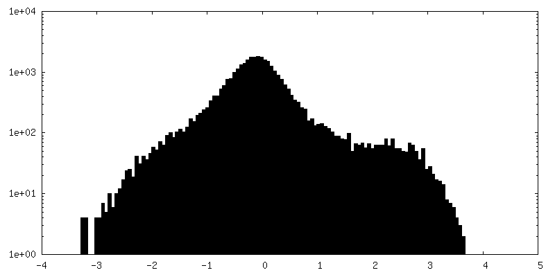
| Density Histograms |


-Half map: Half map 1 from refinement.
| File | emd_17467_half_map_1.map | ||||||||||||
|---|---|---|---|---|---|---|---|---|---|---|---|---|---|
| Annotation | Half map 1 from refinement. | ||||||||||||
| Projections & Slices |
| ||||||||||||
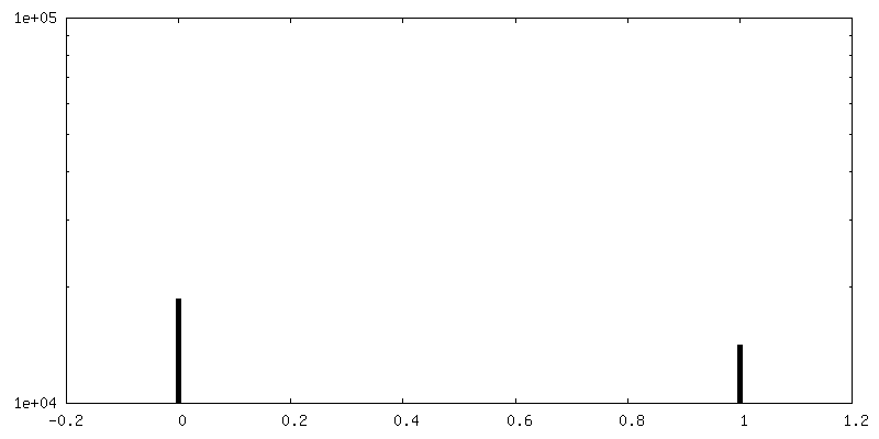
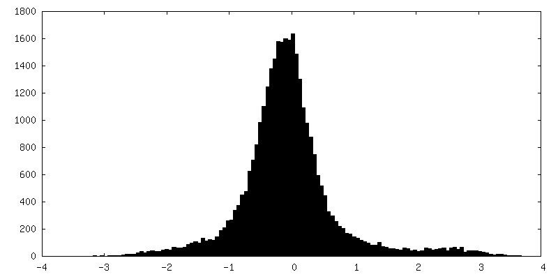
| Density Histograms |


-Half map: Half map 1 from refinement.
| File | emd_17467_half_map_2.map | ||||||||||||
|---|---|---|---|---|---|---|---|---|---|---|---|---|---|
| Annotation | Half map 1 from refinement. | ||||||||||||
| Projections & Slices |
| ||||||||||||
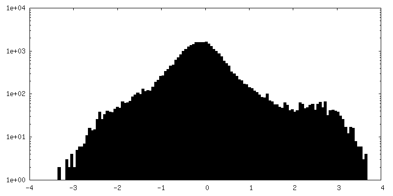
| Density Histograms |


- Sample components
-Entire : Subtomogram representing a segment of the outer wall of cargo-fil...
| Entire | Name: Subtomogram representing a segment of the outer wall of cargo-filled, germinated polar tubes from Vairimorpha necatrix. |
|---|---|
| Components |
|
-Supramolecule #1: Subtomogram representing a segment of the outer wall of cargo-fil...
| Supramolecule | Name: Subtomogram representing a segment of the outer wall of cargo-filled, germinated polar tubes from Vairimorpha necatrix. type: organelle_or_cellular_component / ID: 1 / Parent: 0 |
|---|---|
| Source (natural) | Organism: Vairimorpha necatrix (fungus) / Organ: Polar tube / Location in cell: spores |
-Experimental details
-Structure determination
| Method | cryo EM |
|---|---|
 Processing Processing | subtomogram averaging |
| Aggregation state | filament |
-Sample preparation
| Buffer | pH: 8 Component:
Details: Germination buffer (0.17 mM KCl, 1 mM Tris-HCl (pH 8.0), 10 mM EDTA) | ||||||||||||
|---|---|---|---|---|---|---|---|---|---|---|---|---|---|
| Grid | Model: PELCO Ultrathin Carbon with Lacey Carbon / Material: COPPER / Mesh: 200 / Support film - Material: CARBON / Support film - topology: LACEY / Support film - Film thickness: 200 | ||||||||||||
| Vitrification | Cryogen name: ETHANE / Chamber humidity: 100 % / Chamber temperature: 277.15 K / Instrument: FEI VITROBOT MARK IV | ||||||||||||
| Details | Subtomogram representing a segment of the outer wall of cargo-filled, germinated polar tubes from Vairimorpha nectrix. |
- Electron microscopy
| Microscope | FEI TITAN KRIOS |
|---|---|
| Image recording | Film or detector model: GATAN K2 QUANTUM (4k x 4k) / Detector mode: COUNTING / Average exposure time: 1.0 sec. / Average electron dose: 2.0 e/Å2 |
| Electron beam | Acceleration voltage: 300 kV / Electron source:  FIELD EMISSION GUN FIELD EMISSION GUN |
| Electron optics | Illumination mode: FLOOD BEAM / Imaging mode: OTHER / Nominal defocus max: 5.0 µm / Nominal defocus min: 1.5 µm |
| Experimental equipment |  Model: Titan Krios / Image courtesy: FEI Company |
+Image processing
-Atomic model buiding 1
| Refinement | Protocol: OTHER |
|---|
