 ムービー
ムービー コントローラー
 構造ビューア
 EMN検索について


-検索条件
-検索結果
検索 (著者・登録者: arkin & m)の結果94件中、1から50件目までを表示しています

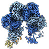
PDB-8c5v: 
Chemotaxis core signalling unit from E protein lysed E. coli cells

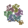
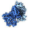
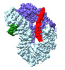
EMDB-28982:
Cryo-EM structure of p97:UBXD1 closed state
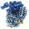
EMDB-28983:
Cryo-EM structure of p97:UBXD1 open state (composite)

EMDB-28987:
Cryo-EM structure of p97:UBXD1 VIM-only state

EMDB-28988:
Cryo-EM structure of p97:UBXD1 meta state
EMDB-28989:
Cryo-EM structure of p97:UBXD1 para state

EMDB-28990:
Cryo-EM structure of p97:UBXD1 PUB-in state
EMDB-28991:
Cryo-EM structure of p97:UBXD1 H4-bound state
EMDB-28992:
Cryo-EM structure of p97:UBXD1 lariat mutant

PDB-8fcl:
Cryo-EM structure of p97:UBXD1 closed state

PDB-8fcm:
Cryo-EM structure of p97:UBXD1 open state
PDB-8fcn:
Cryo-EM structure of p97:UBXD1 VIM-only state
PDB-8fco:
Cryo-EM structure of p97:UBXD1 meta state

PDB-8fcp:
Cryo-EM structure of p97:UBXD1 para state
PDB-8fcq:
Cryo-EM structure of p97:UBXD1 PUB-in state

PDB-8fcr:
Cryo-EM structure of p97:UBXD1 H4-bound state

PDB-8fct:
Cryo-EM structure of p97:UBXD1 lariat mutant
EMDB-28984:
Cryo-EM structure of p97:UBXD1 open state (consensus)
EMDB-28985:
Focused map of p97:UBXD1 open state (P1 protomer)
EMDB-28986:
Focused map of p97:UBXD1 open state (P6 protomer)

EMDB-28756:
Structure of SARS-CoV-2 Omicron BA.1 spike in complex with antibody Fab 1C3

EMDB-28757:
Structure of SARS-CoV-2 spike with antibody Fabs 2A10 and 1H2 (Local refinement of the RBD and Fabs 1H2 and 2A10)

EMDB-28763:
Negative stain EM map of SARS-CoV-2 D614G Spike in complex with 2A10 IgG

EMDB-28764:
Negative stain EM map of SARS-CoV-2 D614G Spike in complex with 4H4 IgG

EMDB-28765:
Negative stain EM map of SARS-CoV-2 D614G Spike in complex with 1C3 IgG

EMDB-28769:
Negative stain EM map of SARS-CoV-2 Omicron BA.1 Spike in complex with 1C3 IgG

EMDB-28770:
Negative stain EM map of SARS-CoV-2 D614G Spike in complex with 2G3 IgG

EMDB-28771:
Negative stain EM map of SARS-CoV-2 D614G Spike in complex with 2E6 IgG

EMDB-28772:
Negative stain EM map of SARS-CoV-2 D614G Spike in complex with 1H2 Fab

EMDB-28773:
Negative stain EM map of SARS-CoV-2 D614G Spike in complex with 1G8 IgG

PDB-8f0g:
Structure of SARS-CoV-2 Omicron BA.1 spike in complex with antibody Fab 1C3

PDB-8f0h:
Structure of SARS-CoV-2 spike with antibody Fabs 2A10 and 1H2 (Local refinement of the RBD and Fabs 1H2 and 2A10)
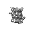
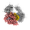
EMDB-17118:
20S proteasome & CBR3 complex
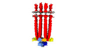
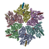
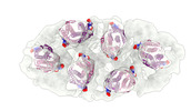
EMDB-15641:
Structure of the Native Chemotaxis Core Signalling Complex from E-gene lysed E. coli cells.

EMDB-15642:
Native Chemotaxis Core Signalling Complex from E. coli, with focused alignment on the baseplate (CheA-CheW)
EMDB-15643:
Native Chemotaxis Core Signalling Complex from E. coli, Focused alignment on ligand binding domain
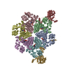
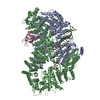
EMDB-14694:
Structure of ubiquitinated FANCI in complex with FANCD2 and double-stranded DNA
PDB-7zf1:
Structure of ubiquitinated FANCI in complex with FANCD2 and double-stranded DNA

EMDB-14719:
FANCD2 Middle and C-terminal domains with USP1-NTE (focused refinement)

EMDB-26653:
Native Lassa glycoprotein in complex with neutralizing antibodies 8.9F and 37.2D

EMDB-26655:
Native Lassa glycoprotein in complex with neutralizing antibodies 12.1F and 37.2D

PDB-7uot:
Native Lassa glycoprotein in complex with neutralizing antibodies 8.9F and 37.2D

PDB-7uov:
Native Lassa glycoprotein in complex with neutralizing antibodies 12.1F and 37.2D

EMDB-14720:
USP1 bound to ubiquitin conjugated to FANCD2 (focused refinement)
EMDB-14721:
USP1 bound to ML323 and ubiquitin conjugated to FANCD2 (focused refinement)

EMDB-14722:
Cryo-EM structure of USP1-UAF1 bound to FANCI and mono-ubiquitinated FANCD2 with ML323 (consensus reconstruction)
EMDB-15284:
Cryo-EM structure of USP1-UAF1 bound to FANCI and mono-ubiquitinated FANCD2 without ML323 (consensus reconstruction)

PDB-7zh3:
USP1 bound to ubiquitin conjugated to FANCD2 (focused refinement)

PDB-7zh4:
USP1 bound to ML323 and ubiquitin conjugated to FANCD2 (focused refinement)

PDB-8a9j:
Cryo-EM structure of USP1-UAF1 bound to FANCI and mono-ubiquitinated FANCD2 without ML323 (consensus reconstruction)
ページ:
 wwPDBはEMDBデータモデルのバージョン3へ移行します
