1P27
 
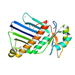
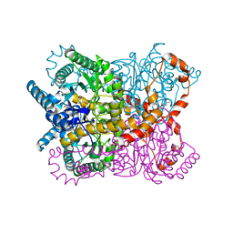
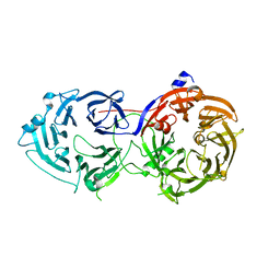
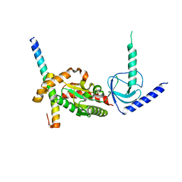
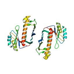 | | Crystal Structure of the Human Y14/Magoh complex | | Descriptor: | Mago nashi protein homolog, RNA-binding protein 8A | | Authors: | Lau, C.K, Diem, M.D, Dreyfuss, G, Van Duyne, G.D. | | Deposit date: | 2003-04-14 | | Release date: | 2003-08-19 | | Last modified: | 2024-02-14 | | Method: | X-RAY DIFFRACTION (2 Å) | | Cite: | Structure of the y14-magoh core of the exon junction complex.
Curr.Biol., 13, 2003
|
|
2X1G
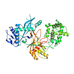
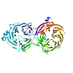
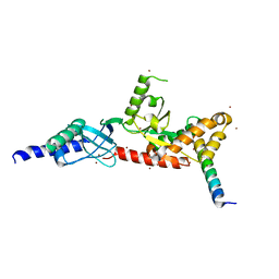
 | | Crystal structure of Importin13 - Mago-Y14 complex | | Descriptor: | CADMUS, PROTEIN MAGO NASHI, RNA-BINDING PROTEIN 8A | | Authors: | Bono, F, Cook, A.G, Gruenwald, M, Ebert, J, Conti, E. | | Deposit date: | 2009-12-23 | | Release date: | 2010-02-16 | | Last modified: | 2023-12-20 | | Method: | X-RAY DIFFRACTION (3.35 Å) | | Cite: | Nuclear Import Mechanism of the Ejc Component Mago- Y14 Revealed by Structural Studies of Importin 13.
Mol.Cell, 37, 2010
|
|
1HL6
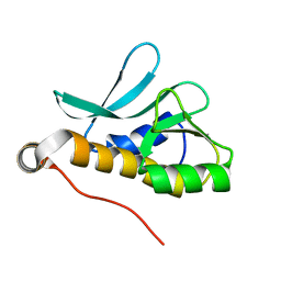
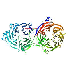
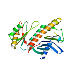 | | A novel mode of RBD-protein recognition in the Y14-mago complex | | Descriptor: | CG8781 PROTEIN, MAGO NASHI PROTEIN | | Authors: | Fribourg, S, Gatfield, D, Yao, W, Izaurralde, E, Conti, E. | | Deposit date: | 2003-03-13 | | Release date: | 2003-05-08 | | Last modified: | 2024-05-08 | | Method: | X-RAY DIFFRACTION (2.5 Å) | | Cite: | A Novel Mode of Rbd-Protein Recognition in the Y14-Mago Complex
Nat.Struct.Biol., 10, 2003
|
|
1RK8
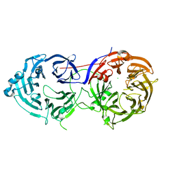
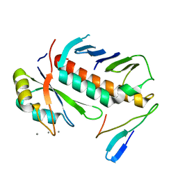 | | Structure of the cytosolic protein PYM bound to the Mago-Y14 core of the exon junction complex | | Descriptor: | CALCIUM ION, CG8781-PA protein, Mago nashi protein, ... | | Authors: | Bono, F, Ebert, J, Guettler, T, Izaurralde, E, Conti, E. | | Deposit date: | 2003-11-21 | | Release date: | 2004-04-13 | | Last modified: | 2024-02-14 | | Method: | X-RAY DIFFRACTION (1.9 Å) | | Cite: | Molecular insights into the interaction of PYM with
the Mago-Y14 core of the exon junction complex
EMBO Rep., 5, 2004
|
|
1OO0
 | |
8XHS
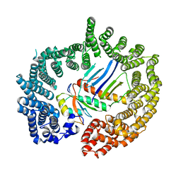
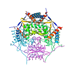
 | | Cryo-EM structure of free-state KmAgo | | Descriptor: | KmAgo | | Authors: | Tao, X, Ding, H, Wu, S. | | Deposit date: | 2023-12-18 | | Release date: | 2024-12-25 | | Last modified: | 2025-07-16 | | Method: | ELECTRON MICROSCOPY (2.85 Å) | | Cite: | Structural and mechanistic insights into a mesophilic prokaryotic Argonaute.
Nucleic Acids Res., 52, 2024
|
|
2XB2
 | | Crystal structure of the core Mago-Y14-eIF4AIII-Barentsz-UPF3b assembly shows how the EJC is bridged to the NMD machinery | | Descriptor: | EUKARYOTIC INITIATION FACTOR 4A-III, MAGNESIUM ION, PHOSPHOAMINOPHOSPHONIC ACID-ADENYLATE ESTER, ... | | Authors: | Buchwald, G, Ebert, J, Basquin, C, Sauliere, J, Jayachandran, U, Bono, F, Le Hir, H, Conti, E. | | Deposit date: | 2010-04-03 | | Release date: | 2010-05-12 | | Last modified: | 2023-12-20 | | Method: | X-RAY DIFFRACTION (3.4 Å) | | Cite: | Insights Into the Recruitment of the Nmd Machinery from the Crystal Structure of a Core Ejc-Upf3B Complex.
Proc.Natl.Acad.Sci.USA, 107, 2010
|
|
8XK0
 | | Structure of the Argonaute protein from Kurthia massiliensis in complex with guide DNA and 19-mer target DNA in pre-cleavage state | | Descriptor: | KmAgo, MANGANESE (II) ION, guide DNA, ... | | Authors: | Tao, X, Ding, H, Wu, S. | | Deposit date: | 2023-12-22 | | Release date: | 2024-12-18 | | Last modified: | 2025-07-16 | | Method: | ELECTRON MICROSCOPY (2.96 Å) | | Cite: | Structural and mechanistic insights into a mesophilic prokaryotic Argonaute.
Nucleic Acids Res., 52, 2024
|
|
8XJW
 | | Structure of the Argonaute protein from Kurthia massiliensis in complex with guide DNA and 19-mer target RNA | | Descriptor: | KmAgo, MANGANESE (II) ION, guide DNA, ... | | Authors: | Tao, X, Ding, H, Wu, S. | | Deposit date: | 2023-12-22 | | Release date: | 2024-12-18 | | Last modified: | 2025-07-16 | | Method: | ELECTRON MICROSCOPY (2.84 Å) | | Cite: | Structural and mechanistic insights into a mesophilic prokaryotic Argonaute.
Nucleic Acids Res., 52, 2024
|
|
8XHV
 | |
8XJX
 | | Structure of the Argonaute protein from Kurthia massiliensis in complex with guide DNA and 19-mer target DNA | | Descriptor: | KmAgo, MANGANESE (II) ION, guide DNA, ... | | Authors: | Tao, X, Ding, H, Wu, S. | | Deposit date: | 2023-12-22 | | Release date: | 2025-01-01 | | Last modified: | 2025-07-16 | | Method: | ELECTRON MICROSCOPY (2.78 Å) | | Cite: | Structural and mechanistic insights into a mesophilic prokaryotic Argonaute.
Nucleic Acids Res., 52, 2024
|
|
8XK3
 | | Structure of the Argonaute protein from Kurthia massiliensis in complex with guide DNA and 19-mer target DNA in target-cleaved state | | Descriptor: | KmAgo, MANGANESE (II) ION, guide DNA, ... | | Authors: | Tao, X, Ding, H, Wu, S. | | Deposit date: | 2023-12-22 | | Release date: | 2025-01-01 | | Last modified: | 2025-07-16 | | Method: | ELECTRON MICROSCOPY (2.76 Å) | | Cite: | Structural and mechanistic insights into a mesophilic prokaryotic Argonaute.
Nucleic Acids Res., 52, 2024
|
|
1OAD
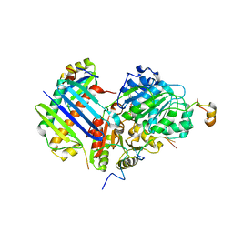
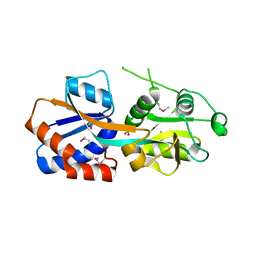
 | | Glucose isomerase from Streptomyces rubiginosus in P21212 crystal form | | Descriptor: | (4R)-2-METHYLPENTANE-2,4-DIOL, (4S)-2-METHYL-2,4-PENTANEDIOL, 2-AMINO-2-HYDROXYMETHYL-PROPANE-1,3-DIOL, ... | | Authors: | Ramagopal, U.A, Dauter, M, Dauter, Z. | | Deposit date: | 2003-01-08 | | Release date: | 2003-01-30 | | Last modified: | 2024-05-08 | | Method: | X-RAY DIFFRACTION (1.5 Å) | | Cite: | Sad Manganese in Two Crystal Forms of Glucose Isomerase
Acta Crystallogr.,Sect.D, 59, 2003
|
|
1TWY
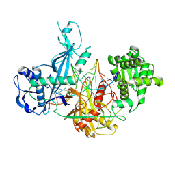
 | | Crystal structure of an ABC-type phosphate transport receptor from Vibrio cholerae | | Descriptor: | ABC transporter, periplasmic substrate-binding protein, MAGNESIUM ION, ... | | Authors: | Ramagopal, U.A, Patskovsky, Y, Almo, S.C, Burley, S.K, New York SGX Research Center for Structural Genomics (NYSGXRC) | | Deposit date: | 2004-07-01 | | Release date: | 2004-12-21 | | Last modified: | 2024-10-16 | | Method: | X-RAY DIFFRACTION (1.65 Å) | | Cite: | Crystal structure of hypothetical ABC-type phosphate transporter
To be Published
|
|
2A1V
 | |
2A2L
 | |
6N8S
 | |
6N8R
 | |
6N8Q
 | |
6N8P
 | |
4DEX
 | |
4DEY
 | |
5TRU
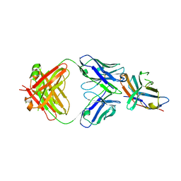 | | Structure of the first-in-class checkpoint inhibitor Ipilimumab bound to human CTLA-4 | | Descriptor: | Cytotoxic T-lymphocyte protein 4, Ipilimumab Fab heavy chain, Ipilimumab Fab light chain | | Authors: | Ramagopal, U.A, Liu, W, Garrett-Thomson, S.C, Yan, Q, Srinivasan, M, Wong, S.C, Bell, A, Mankikar, S, Rangan, V.S, Deshpande, S, Bonanno, J.B, Korman, A.J, Almo, S.C. | | Deposit date: | 2016-10-27 | | Release date: | 2017-05-10 | | Last modified: | 2024-11-20 | | Method: | X-RAY DIFFRACTION (3 Å) | | Cite: | Structural basis for cancer immunotherapy by the first-in-class checkpoint inhibitor ipilimumab.
Proc. Natl. Acad. Sci. U.S.A., 114, 2017
|
|
6FMG
 | |
4ETO
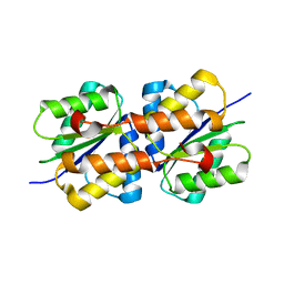
 | | Structure of S100A4 in complex with non-muscle myosin-IIA peptide | | Descriptor: | CALCIUM ION, Myosin-9, Protein S100-A4 | | Authors: | Ramagopal, U.A, Dulyaninova, N.G, Kumar, P.R, Almo, S.C, Bresnick, A.R, New York Structural Genomics Research Consortium (NYSGRC), Atoms-to-Animals: The Immune Function Network (IFN) | | Deposit date: | 2012-04-24 | | Release date: | 2012-07-11 | | Last modified: | 2023-09-13 | | Method: | X-RAY DIFFRACTION (1.54 Å) | | Cite: | Structure of S100A4 with bound peptide P
To be Published
|
|


