Loading
PDBj
MenuPDBj@FacebookPDBj@TwitterPDBj@YouTubewwPDB FoundationwwPDB
RCSB PDBPDBeBMRBAdv. SearchSearch help

4UOW
DownloadVisualize
BU of 4uow by Molmil
Crystal structure of the titin M10-Obscurin Ig domain 1 complex
Descriptor: CHLORIDE ION, Obscurin, SODIUM ION, ...
Authors:Pernigo, S, Fukuzawa, A, Gautel, M, Steiner, R.A.
Deposit date:2014-06-10
Release date:2014-12-17
Last modified:2024-01-10
Method:X-RAY DIFFRACTION (3.3 Å)
Cite:The Crystal Structure of the Human Titin:Obscurin Complex Reveals a Conserved Yet Specific Muscle M-Band Zipper Module.
J.Mol.Biol., 427, 2015
1IE5
DownloadVisualize
BU of 1ie5 by Molmil
NMR STRUCTURE OF THE THIRD IMMUNOGLOBULIN DOMAIN FROM THE NEURAL CELL ADHESION MOLECULE.
Descriptor: NEURAL CELL ADHESION MOLECULE
Authors:Atkins, A.R, Chung, J, Deechongkit, S, Little, E.B, Edelman, G.M, Wright, P.E, Cunningham, B.A, Dyson, H.J.
Deposit date:2001-04-06
Release date:2001-08-08
Last modified:2022-02-23
Method:SOLUTION NMR
Cite:Solution structure of the third immunoglobulin domain of the neural cell adhesion molecule N-CAM: can solution studies define the mechanism of homophilic binding?
J.Mol.Biol., 311, 2001
6NRX
DownloadVisualize
BU of 6nrx by Molmil
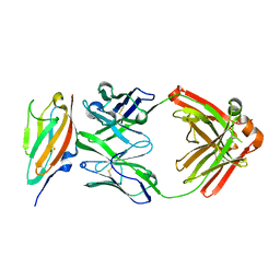
Crystal structure of DIP-eta IG1 homodimer
Descriptor: 2-acetamido-2-deoxy-beta-D-glucopyranose, Dpr-interacting protein eta, isoform B, ...
Authors:Cheng, S, Park, Y.J, Kurleto, J.D, Ozkan, E.
Deposit date:2019-01-24
Release date:2019-02-06
Last modified:2023-10-11
Method:X-RAY DIFFRACTION (1.9 Å)
Cite:Molecular basis of synaptic specificity by immunoglobulin superfamily receptors in Drosophila.
Elife, 8, 2019
5AEA
DownloadVisualize
BU of 5aea by Molmil
Crystal structure of human NCAM domain 1
Descriptor: CITRATE ANION, NEURAL CELL ADHESION MOLECULE 1
Authors:Kvansakul, M, Griffiths, K, Foley, M.
Deposit date:2015-08-27
Release date:2016-04-13
Last modified:2024-01-10
Method:X-RAY DIFFRACTION (1.9 Å)
Cite:I-Bodies, Human Single Domain Antibodies that Antagonize Chemokine Receptor Cxcr4.
J.Biol.Chem., 291, 2016
6NRQ
DownloadVisualize
BU of 6nrq by Molmil
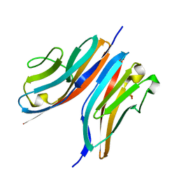
Crystal structure of Dpr10 IG1 bound to DIP-alpha IG1
Descriptor: 2-acetamido-2-deoxy-beta-D-glucopyranose, Defective proboscis extension response 10, isoform A, ...
Authors:Cheng, S, Park, Y.J, Kurleto, J.D, Ozkan, E.
Deposit date:2019-01-24
Release date:2019-02-06
Last modified:2023-10-11
Method:X-RAY DIFFRACTION (1.8 Å)
Cite:Molecular basis of synaptic specificity by immunoglobulin superfamily receptors in Drosophila.
Elife, 8, 2019
8OVU
DownloadVisualize
BU of 8ovu by Molmil
Human titin immunoglobulin-like 21 domain
Descriptor: MAGNESIUM ION, Titin
Authors:Martinez-Martin, I, Crousilles, A, Mortensen, S.A, Alegre-Cebollada, J, Wilmanns, M.
Deposit date:2023-04-26
Release date:2023-11-29
Last modified:2024-01-10
Method:X-RAY DIFFRACTION (1.95 Å)
Cite:Titin domains with reduced core hydrophobicity cause dilated cardiomyopathy.
Cell Rep, 42, 2023
8P35
DownloadVisualize
BU of 8p35 by Molmil
Mutant human titin immunoglobulin-like 21 domain - C3575S
Descriptor: Titin
Authors:Martinez-Martin, I, Crousilles, A, Mortensen, S.A, Alegre-Cebollada, J, Wilmanns, M.
Deposit date:2023-05-17
Release date:2023-11-29
Last modified:2024-01-10
Method:X-RAY DIFFRACTION (2.2 Å)
Cite:Titin domains with reduced core hydrophobicity cause dilated cardiomyopathy.
Cell Rep, 42, 2023
7Y95
DownloadVisualize
BU of 7y95 by Molmil
Crystal structure of sDscam Ig1 domain, isoform beta6v2
Descriptor: Dscam, GLYCEROL, SODIUM ION
Authors:Chen, Q, Yu, Y, Cheng, J.
Deposit date:2022-06-24
Release date:2023-05-24
Last modified:2023-11-29
Method:X-RAY DIFFRACTION (1.55 Å)
Cite:Structural basis for the self-recognition of sDSCAM in Chelicerata.
Nat Commun, 14, 2023
4CBP
DownloadVisualize
BU of 4cbp by Molmil
Crystal structure of neural ectodermal development factor IMP-L2.
Descriptor: GLYCEROL, NEURAL/ECTODERMAL DEVELOPMENT FACTOR IMP-L2
Authors:Kulahin, N, Kristensen, O, Brzozowski, M, Schluckebier, G, Meyts, P.D.
Deposit date:2013-10-15
Release date:2014-10-29
Last modified:2019-05-29
Method:X-RAY DIFFRACTION (1.6 Å)
Cite:Structural Analysis of Imp-L2 Function
To be Published
4C4K
DownloadVisualize
BU of 4c4k by Molmil
Crystal structure of the titin M10-Obscurin Ig domain 1 complex
Descriptor: 1,2-ETHANEDIOL, OBSCURIN, TITIN
Authors:Pernigo, S, Fukuzawa, A, Gautel, M, Steiner, R.A.
Deposit date:2013-09-05
Release date:2014-09-24
Last modified:2023-12-20
Method:X-RAY DIFFRACTION (1.95 Å)
Cite:The Crystal Structure of the Human Titin:Obscurin Complex Reveals a Conserved Yet Specific Muscle M-Band Zipper Module.
J.Mol.Biol., 427, 2015
3DAR
DownloadVisualize
BU of 3dar by Molmil
Crystal structure of D2 domain from human FGFR2
Descriptor: Fibroblast growth factor receptor 2
Authors:Brown, A, Blundell, T.L.
Deposit date:2008-05-30
Release date:2008-06-10
Last modified:2023-08-30
Method:X-RAY DIFFRACTION (2.2 Å)
Cite:Crystal structure of the FGFR2 D2 domain
To be Published
3CAF
DownloadVisualize
BU of 3caf by Molmil
Crystal Structure of hFGFR2 D2 Domain
Descriptor: Fibroblast growth factor receptor 2
Authors:Guo, F, Dakshinamurthy, R, Kathir, K.M, Thallapuranam, S.K.K, Sakon, J.
Deposit date:2008-02-19
Release date:2009-01-27
Last modified:2011-07-13
Method:X-RAY DIFFRACTION (1.96 Å)
Cite:Crystal structure analysis of the hFGFR2 D2 domain
To be Published
6FWX
DownloadVisualize
BU of 6fwx by Molmil
Chimeric titin Z1Z2-Z1Z2 tandem (Z1212) functionalized with a GRGDS exogenous peptide from fibronectin
Descriptor: 1,2-ETHANEDIOL, Titin,Titin,Titin
Authors:Mayans, O, Fleming, J, Hill, C.
Deposit date:2018-03-07
Release date:2019-03-27
Last modified:2024-01-17
Method:X-RAY DIFFRACTION (3 Å)
Cite:Self-Assembling Proteins as High-Performance Substrates for Embryonic Stem Cell Self-Renewal.
Adv. Mater. Weinheim, 31, 2019
3CX2
DownloadVisualize
BU of 3cx2 by Molmil
Crystal structure of the C1 domain of cardiac isoform of myosin binding protein-C at 1.3A
Descriptor: Myosin-binding protein C, cardiac-type
Authors:Fisher, S.J, Helliwell, J.R, Khurshid, S, Govada, L, Redwood, C, Squire, J.M, Chayen, N.E.
Deposit date:2008-04-23
Release date:2008-07-01
Last modified:2023-08-30
Method:X-RAY DIFFRACTION (1.3 Å)
Cite:An investigation into the protonation states of the C1 domain of cardiac myosin-binding protein C
Acta Crystallogr.,Sect.D, 64, 2008
6HCI
DownloadVisualize
BU of 6hci by Molmil
Crystal structure of titin M3 domain
Descriptor: SULFATE ION, Titin
Authors:Chatziefthimiou, S.D, Ugurlar, D, Wilmanns, M.
Deposit date:2018-08-15
Release date:2019-08-28
Last modified:2024-05-01
Method:X-RAY DIFFRACTION (2.12 Å)
Cite:Structural diversity in the atomic resolution 3D fingerprint of the titin M-band segment.
Plos One, 14, 2019
6H4L
DownloadVisualize
BU of 6h4l by Molmil
Structure of Titin M4 trigonal form
Descriptor: CHLORIDE ION, Titin, ZINC ION
Authors:Sauer, F, Wilmanns, M.
Deposit date:2018-07-21
Release date:2019-08-07
Last modified:2020-02-19
Method:X-RAY DIFFRACTION (1.6 Å)
Cite:Structural diversity in the atomic resolution 3D fingerprint of the titin M-band segment.
Plos One, 14, 2019
3EUU
DownloadVisualize
BU of 3euu by Molmil
Crystal structure of the FGFR2 D2 domain
Descriptor: Fibroblast growth factor receptor 2
Authors:Sakon, J, Guo, F, Dakshinamurthy, R, Kathir, K.M, Thallapuranam, S.K.K.
Deposit date:2008-10-10
Release date:2009-08-25
Last modified:2023-09-06
Method:X-RAY DIFFRACTION (2.34 Å)
Cite:Crystal structure of the D2 domain of hFGFR2
To be Published
1TNN
DownloadVisualize
BU of 1tnn by Molmil
Tertiary structure of an immunoglobulin-like domain from the giant muscle protein titin: a new member of the I set
Descriptor: TITIN MODULE M5
Authors:Pfuhl, M, Pastore, A.
Deposit date:1995-01-17
Release date:1995-04-20
Last modified:2024-05-01
Method:SOLUTION NMR
Cite:Tertiary structure of an immunoglobulin-like domain from the giant muscle protein titin: a new member of the I set.
Structure, 3, 1995
1U2H
DownloadVisualize
BU of 1u2h by Molmil
X-ray Structure of the N-terminally truncated human APEP-1
Descriptor: Aortic preferentially expressed protein 1
Authors:Manjasetty, B.A, Scheich, C, Roske, Y, Niesen, F.H, Gotz, F, Bussow, K, Heinemann, U.
Deposit date:2004-07-19
Release date:2005-07-05
Last modified:2023-10-25
Method:X-RAY DIFFRACTION (0.96 Å)
Cite:X-ray structure of engineered human Aortic Preferentially Expressed Protein-1 (APEG-1)
Bmc Struct.Biol., 5, 2005
1RHF
DownloadVisualize
BU of 1rhf by Molmil
Crystal Structure of human Tyro3-D1D2
Descriptor: 4-(2-HYDROXYETHYL)-1-PIPERAZINE ETHANESULFONIC ACID, ACETATE ION, Tyrosine-protein kinase receptor TYRO3, ...
Authors:Heiring, C, Dahlback, B, Muller, Y.A.
Deposit date:2003-11-14
Release date:2004-03-23
Last modified:2017-10-11
Method:X-RAY DIFFRACTION (1.96 Å)
Cite:Ligand recognition and homophilic interactions in Tyro3: structural insights into the Axl/Tyro3 receptor tyrosine kinase family.
J.Biol.Chem., 279, 2004
1TNM
DownloadVisualize
BU of 1tnm by Molmil
TERTIARY STRUCTURE OF AN IMMUNOGLOBULIN-LIKE DOMAIN FROM THE GIANT MUSCLE PROTEIN TITIN: A NEW MEMBER OF THE I SET
Descriptor: TITIN MODULE M5
Authors:Pfuhl, M, Pastore, A.
Deposit date:1995-01-17
Release date:1995-04-20
Last modified:2024-05-01
Method:SOLUTION NMR
Cite:Tertiary structure of an immunoglobulin-like domain from the giant muscle protein titin: a new member of the I set.
Structure, 3, 1995
1TLK
DownloadVisualize
BU of 1tlk by Molmil
X-RAY STRUCTURE DETERMINATION OF TELOKIN, THE C-TERMINAL DOMAIN OF MYOSIN LIGHT CHAIN KINASE, AT 2.8 ANGSTROMS RESOLUTION
Descriptor: TELOKIN
Authors:Holden, H.M, Rayment, I.
Deposit date:1992-07-20
Release date:1993-10-31
Last modified:2024-02-14
Method:X-RAY DIFFRACTION (2.8 Å)
Cite:X-ray structure determination of telokin, the C-terminal domain of myosin light chain kinase, at 2.8 A resolution.
J.Mol.Biol., 227, 1992
2LQR
DownloadVisualize
BU of 2lqr by Molmil
NMR structure of Ig3 domain of palladin
Descriptor: Palladin
Authors:Beck, M.R, Dixon IV, R.D.S, Otey, C.A, Campbell, S.L, Murphy, G.S.
Deposit date:2012-03-13
Release date:2013-01-23
Last modified:2024-05-01
Method:SOLUTION NMR
Cite:Structure and Function of Palladin's Actin Binding Domain.
J.Mol.Biol., 425, 2013
1WWB
DownloadVisualize
BU of 1wwb by Molmil
LIGAND BINDING DOMAIN OF HUMAN TRKB RECEPTOR
Descriptor: PROTEIN (Brain Derived Neurotrophic Factor Receptor TrkB)
Authors:Wiesmann, C, Ultsch, M.H, Bass, S.H, De Vos, A.M.
Deposit date:1999-05-03
Release date:1999-07-07
Last modified:2023-08-23
Method:X-RAY DIFFRACTION (2.1 Å)
Cite:Crystal structures of the neurotrophin-binding domain of TrkA, TrkB and TrkC.
J.Mol.Biol., 290, 1999
1WVZ
DownloadVisualize
BU of 1wvz by Molmil
Solution Structure of the D2 Domain of the Fibroblast Growth Factor
Descriptor: Fibroblast growth factor receptor 2
Authors:Hung, K.W, Kumar, T.K.S, Yu, C.
Deposit date:2004-12-28
Release date:2005-12-28
Last modified:2022-03-02
Method:SOLUTION NMR
Cite:Solution Structure of the D2 Domain of the Fibroblast Growth Factor
To be Published

225158

건을2024-09-18부터공개중

PDB statisticsPDBj update infoContact PDBjnumon