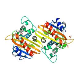
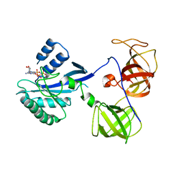
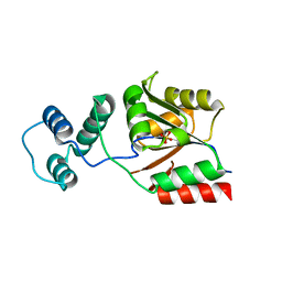
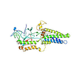
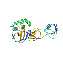
1UYJ
 
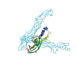 | | Clostridium perfringens epsilon toxin shows structural similarity with the pore forming toxin aerolysin | | 分子名称: | EPSILON-TOXIN, URANIUM ATOM | | 著者 | Cole, A.R, Gibert, M, Poppoff, M, Moss, D.S, Titball, R.W, Basak, A.K. | | 登録日 | 2004-03-02 | | 公開日 | 2004-08-05 | | 最終更新日 | 2024-05-08 | | 実験手法 | X-RAY DIFFRACTION (2.6 Å) | | 主引用文献 | Clostridium Perfringens Epsilon-Toxin Shows Structural Similarity to the Pore-Forming Toxin Aerolysin
Nat.Struct.Mol.Biol., 11, 2004
|
|
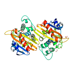
1PFO
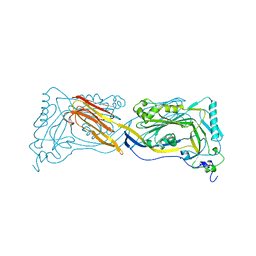 | | PERFRINGOLYSIN O | | 分子名称: | PERFRINGOLYSIN O | | 著者 | Rossjohn, J, Parker, M.W. | | 登録日 | 1997-07-31 | | 公開日 | 1998-08-05 | | 最終更新日 | 2024-02-14 | | 実験手法 | X-RAY DIFFRACTION (2.2 Å) | | 主引用文献 | Structure of a cholesterol-binding, thiol-activated cytolysin and a model of its membrane form.
Cell(Cambridge,Mass.), 89, 1997
|
|
2WGW
 | | Crystal structure of the OXA-10 V117T mutant at pH 8.0 | | 分子名称: | BETA-LACTAMASE OXA-10, GLYCEROL, SULFATE ION | | 著者 | Vercheval, L, Kerff, F, Bauvois, C, Sauvage, E, Guiet, R, Charlier, P, Galleni, M. | | 登録日 | 2009-04-27 | | 公開日 | 2010-05-19 | | 最終更新日 | 2023-12-13 | | 実験手法 | X-RAY DIFFRACTION (1.8 Å) | | 主引用文献 | Three Factors that Modulate the Activity of Class D Beta-Lactamases and Interfere with the Post-Translational Carboxylation of Lys70.
Biochem.J., 432, 2010
|
|
2WGV
 | | Crystal structure of the OXA-10 V117T mutant at pH 6.5 inhibited by a chloride ion | | 分子名称: | BETA-LACTAMASE OXA-10, CHLORIDE ION, CITRIC ACID, ... | | 著者 | Vercheval, L, Kerff, F, Bauvois, C, Sauvage, E, Guiet, R, Charlier, P, Galleni, M. | | 登録日 | 2009-04-27 | | 公開日 | 2010-05-19 | | 最終更新日 | 2023-12-13 | | 実験手法 | X-RAY DIFFRACTION (1.8 Å) | | 主引用文献 | Three Factors that Modulate the Activity of Class D Beta-Lactamases and Interfere with the Post- Translational Carboxylation of Lys70.
Biochem.J., 432, 2010
|
|
2WKH
 | | Crystal structure of the acyl-enzyme OXA-10 K70C-Ampicillin at pH 7 | | 分子名称: | (2R,4S)-2-[(R)-{[(2R)-2-amino-2-phenylacetyl]amino}(carboxy)methyl]-5,5-dimethyl-1,3-thiazolidine-4-carboxylic acid, BETA-LACTAMASE OXA-10, SULFATE ION | | 著者 | Vercheval, L, Bauvois, C, Kerff, F, Sauvage, E, Guiet, R, Charlier, P, Galleni, M. | | 登録日 | 2009-06-11 | | 公開日 | 2010-08-25 | | 最終更新日 | 2023-12-13 | | 実験手法 | X-RAY DIFFRACTION (1.791 Å) | | 主引用文献 | Three Factors that Modulate the Activity of Class D Beta-Lactamases and Interfere with the Post-Translational Carboxylation of Lys70.
Biochem.J., 432, 2010
|
|
2WKI
 | | Crystal structure of the OXA-10 K70C mutant at pH 7.0 | | 分子名称: | 1,2-ETHANEDIOL, BETA-LACTAMASE OXA-10, GLYCEROL, ... | | 著者 | Vercheval, L, Bauvois, C, Kerff, F, Sauvage, E, Guiet, R, Charlier, P, Galleni, M. | | 登録日 | 2009-06-11 | | 公開日 | 2010-08-25 | | 最終更新日 | 2024-10-09 | | 実験手法 | X-RAY DIFFRACTION (2.1 Å) | | 主引用文献 | Three Factors that Modulate the Activity of Class D Beta-Lactamases and Interfere with the Post-Translational Carboxylation of Lys70.
Biochem.J., 432, 2010
|
|
5WX9
 | | Crystal Structure of AtERF96 with GCC-box | | 分子名称: | Ethylene-responsive transcription factor ERF096, GCC-box motif | | 著者 | Chen, C.Y, Cheng, Y.S. | | 登録日 | 2017-01-06 | | 公開日 | 2017-11-22 | | 最終更新日 | 2023-11-22 | | 実験手法 | X-RAY DIFFRACTION (1.76 Å) | | 主引用文献 | Structural insights into Arabidopsis ethylene response factor 96 with an extended N-terminal binding to GCC box.
Plant Mol.Biol., 104, 2020
|
|
1R5B
 | | Crystal structure analysis of sup35 | | 分子名称: | Eukaryotic peptide chain release factor GTP-binding subunit | | 著者 | Kong, C, Song, H. | | 登録日 | 2003-10-10 | | 公開日 | 2004-05-25 | | 最終更新日 | 2024-03-13 | | 実験手法 | X-RAY DIFFRACTION (2.35 Å) | | 主引用文献 | Crystal structure and functional analysis of the eukaryotic class II release factor eRF3 from S. pombe
Mol.Cell, 14, 2004
|
|
1R5O
 | | crystal structure analysis of sup35 complexed with GMPPNP | | 分子名称: | Eukaryotic peptide chain release factor GTP-binding subunit, PHOSPHOAMINOPHOSPHONIC ACID-GUANYLATE ESTER | | 著者 | Kong, C, Song, H. | | 登録日 | 2003-10-11 | | 公開日 | 2004-05-25 | | 最終更新日 | 2023-10-25 | | 実験手法 | X-RAY DIFFRACTION (3.2 Å) | | 主引用文献 | Crystal structure and functional analysis of the eukaryotic class II release factor eRF3 from S. pombe
Mol.Cell, 14, 2004
|
|
1R5N
 | | Crystal Structure Analysis of sup35 complexed with GDP | | 分子名称: | Eukaryotic peptide chain release factor GTP-binding subunit, GUANOSINE-5'-DIPHOSPHATE | | 著者 | Kong, C, Song, H. | | 登録日 | 2003-10-10 | | 公開日 | 2004-05-25 | | 最終更新日 | 2023-10-25 | | 実験手法 | X-RAY DIFFRACTION (2.9 Å) | | 主引用文献 | Crystal structure and functional analysis of the eukaryotic class II release factor eRF3 from S. pombe
Mol.Cell, 14, 2004
|
|
5UOT
 | |
4X0L
 | | Human haptoglobin-haemoglobin complex | | 分子名称: | CACODYLATE ION, GLYCEROL, Haptoglobin, ... | | 著者 | Lane-Serff, H, MacGregor, P, Lowe, E.D, Carrington, M, Higgins, M.K. | | 登録日 | 2014-11-21 | | 公開日 | 2014-12-24 | | 最終更新日 | 2024-01-10 | | 実験手法 | X-RAY DIFFRACTION (2.05 Å) | | 主引用文献 | Structural basis for ligand and innate immunity factor uptake by the trypanosome haptoglobin-haemoglobin receptor.
Elife, 3, 2014
|
|
3KZX
 | |
8U57
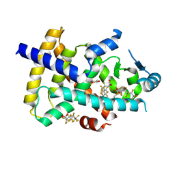 | | PPARg LBD in complex with perfluorooctanoic acid (PFOA) | | 分子名称: | (4S,5S)-1,2-DITHIANE-4,5-DIOL, Peroxisome proliferator-activated receptor gamma, pentadecafluorooctanoic acid | | 著者 | Pederick, J.L, Frkic, R.L, McDougal, D.P, Bruning, J.B. | | 登録日 | 2023-09-12 | | 公開日 | 2024-07-24 | | 実験手法 | X-RAY DIFFRACTION (2 Å) | | 主引用文献 | A structural basis for the activation of peroxisome proliferator-activated receptor gamma (PPAR gamma ) by perfluorooctanoic acid (PFOA).
Chemosphere, 354, 2024
|
|
4X0J
 | | Trypanosoma brucei haptoglobin-haemoglobin receptor | | 分子名称: | Haptoglobin-hemoglobin receptor | | 著者 | Lane-Serff, H, MacGregor, P, Lowe, E.D, Carrington, M, Higgins, M.K. | | 登録日 | 2014-11-21 | | 公開日 | 2014-12-24 | | 最終更新日 | 2024-01-10 | | 実験手法 | X-RAY DIFFRACTION (1.85 Å) | | 主引用文献 | Structural basis for ligand and innate immunity factor uptake by the trypanosome haptoglobin-haemoglobin receptor.
Elife, 3, 2014
|
|
2LGT
 | | Backbone 1H, 13C, and 15N Chemical Shift Assignments for QFM(Y)F | | 分子名称: | Eukaryotic peptide chain release factor subunit 1 | | 著者 | Wong, L.E, Li, Y, Pillay, S, Pervushin, K. | | 登録日 | 2011-08-02 | | 公開日 | 2012-03-14 | | 最終更新日 | 2024-05-01 | | 実験手法 | SOLUTION NMR | | 主引用文献 | Selectivity of stop codon recognition in translation termination is modulated by multiple conformations of GTS loop in eRF1
Nucleic Acids Res., 2012
|
|
8HFC
 | | Cryo-EM structure of yeast Erf2/Erf4 complex | | 分子名称: | PALMITIC ACID, Palmitoyltransferase ERF2, Ras modification protein ERF4, ... | | 著者 | Wu, J, Hu, Q, Zhang, Y, Yang, A, Liu, S. | | 登録日 | 2022-11-10 | | 公開日 | 2023-11-22 | | 最終更新日 | 2024-06-05 | | 実験手法 | ELECTRON MICROSCOPY (3.5 Å) | | 主引用文献 | Regulation of RAS palmitoyltransferases by accessory proteins and palmitoylation.
Nat.Struct.Mol.Biol., 31, 2024
|
|
6SB3
 | |
6SB5
 | |
6OAR
 | |
6OAT
 | |
6D7A
 | | Structure of T. gondii PLP1 beta-rich domain | | 分子名称: | Perforin-like protein 1, SODIUM ION | | 著者 | Guerra, A.J, Koropatkin, N.M, Wawrzak, Z, Bahr, C.M.E, Carruthers, V.B. | | 登録日 | 2018-04-24 | | 公開日 | 2018-05-16 | | 最終更新日 | 2019-12-18 | | 実験手法 | X-RAY DIFFRACTION (1.13 Å) | | 主引用文献 | Structural basis of Toxoplasma gondii perforin-like protein 1 membrane interaction and activity during egress.
PLoS Pathog., 14, 2018
|
|
6SB1
 | | Crystal structure of murine perforin-2 P2 domain crystal form 1 | | 分子名称: | CHLORIDE ION, GLYCEROL, Macrophage-expressed gene 1 protein | | 著者 | Ni, T, Ginger, L, Gilbert, R.J.C. | | 登録日 | 2019-07-18 | | 公開日 | 2020-02-05 | | 最終更新日 | 2020-02-26 | | 実験手法 | X-RAY DIFFRACTION (2.05 Å) | | 主引用文献 | Structure and mechanism of bactericidal mammalian perforin-2, an ancient agent of innate immunity.
Sci Adv, 6, 2020
|
|
6SB4
 | | Crystal structure of murine perforin-2 P2 domain crystal form 2 | | 分子名称: | Macrophage-expressed gene 1 protein | | 著者 | Ni, T, Yu, X, Ginger, L, Gilbert, R.J.C. | | 登録日 | 2019-07-18 | | 公開日 | 2020-02-05 | | 最終更新日 | 2020-02-26 | | 実験手法 | X-RAY DIFFRACTION (3.17 Å) | | 主引用文献 | Structure and mechanism of bactericidal mammalian perforin-2, an ancient agent of innate immunity.
Sci Adv, 6, 2020
|
|
8A1S
 | | Structure of murine perforin-2 (Mpeg1) pore in twisted form | | 分子名称: | 2-acetamido-2-deoxy-beta-D-glucopyranose, Macrophage-expressed gene 1 protein | | 著者 | Yu, X, Ni, T, Zhang, P, Gilbert, R. | | 登録日 | 2022-06-02 | | 公開日 | 2022-07-20 | | 最終更新日 | 2023-10-18 | | 実験手法 | ELECTRON MICROSCOPY (4 Å) | | 主引用文献 | Cryo-EM structures of perforin-2 in isolation and assembled on a membrane suggest a mechanism for pore formation.
Embo J., 41, 2022
|
|


