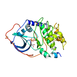
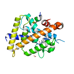
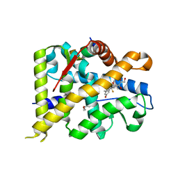
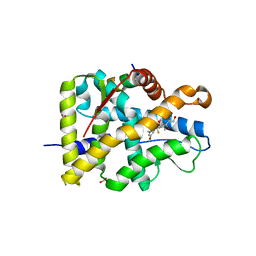
3WTQ
 
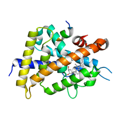 | | Crystal structure of VDR-LBD complexed with 22S-butyl-2-methylidene-19-nor-1a,25-dihydroxyvitamin D3 | | 分子名称: | (1R,3R,7E,17beta)-17-[(2R,3S)-3-butyl-6-hydroxy-6-methylheptan-2-yl]-2-methylidene-9,10-secoestra-5,7-diene-1,3-diol, Mediator of RNA polymerase II transcription subunit 1, Vitamin D3 receptor | | 著者 | Nakabayashi, M, Inaba, Y, Itoh, T, Anami, Y, Ikura, T, Ito, N, Yamamoto, K. | | 登録日 | 2014-04-15 | | 公開日 | 2015-04-15 | | 最終更新日 | 2024-03-20 | | 実験手法 | X-RAY DIFFRACTION (2.1 Å) | | 主引用文献 | Crystal structure of VDR-LBD complexed with 22S-butyl-2-methylidene-19-nor-1a,25-dihydroxyvitamin D3
To be Published
|
|
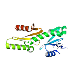
4JST
 | | Structure of Clostridium thermocellum polynucleotide kinase bound to UTP | | 分子名称: | MAGNESIUM ION, Metallophosphoesterase, SODIUM ION, ... | | 著者 | Das, U, Wang, L.K, Smith, P, Shuman, S. | | 登録日 | 2013-03-22 | | 公開日 | 2013-08-28 | | 最終更新日 | 2013-12-18 | | 実験手法 | X-RAY DIFFRACTION (2.03 Å) | | 主引用文献 | Structural and biochemical analysis of the phosphate donor specificity of the polynucleotide kinase component of the bacterial pnkphen1 RNA repair system.
Biochemistry, 52, 2013
|
|
4JSY
 | | Structure of Clostridium thermocellum polynucleotide kinase bound to GTP | | 分子名称: | GUANOSINE-5'-TRIPHOSPHATE, MAGNESIUM ION, Metallophosphoesterase | | 著者 | Das, U, Wang, L.K, Smith, P, Shuman, S. | | 登録日 | 2013-03-22 | | 公開日 | 2013-08-28 | | 最終更新日 | 2013-12-18 | | 実験手法 | X-RAY DIFFRACTION (2.14 Å) | | 主引用文献 | Structural and biochemical analysis of the phosphate donor specificity of the polynucleotide kinase component of the bacterial pnkphen1 RNA repair system.
Biochemistry, 52, 2013
|
|
4JT4
 | | Structure of Clostridium thermocellum polynucleotide kinase bound to dATP | | 分子名称: | 2'-DEOXYADENOSINE 5'-TRIPHOSPHATE, MAGNESIUM ION, Metallophosphoesterase, ... | | 著者 | Das, U, Wang, L.K, Smith, P, Shuman, S. | | 登録日 | 2013-03-22 | | 公開日 | 2013-08-28 | | 最終更新日 | 2013-12-18 | | 実験手法 | X-RAY DIFFRACTION (2.01 Å) | | 主引用文献 | Structural and biochemical analysis of the phosphate donor specificity of the polynucleotide kinase component of the bacterial pnkphen1 RNA repair system.
Biochemistry, 52, 2013
|
|
4JT2
 | | Structure of Clostridium thermocellum polynucleotide kinase bound to CTP | | 分子名称: | CYTIDINE-5'-TRIPHOSPHATE, MAGNESIUM ION, Metallophosphoesterase | | 著者 | Das, U, Wang, L.K, Smith, P, Shuman, S. | | 登録日 | 2013-03-22 | | 公開日 | 2013-08-28 | | 最終更新日 | 2013-12-18 | | 実験手法 | X-RAY DIFFRACTION (2.49 Å) | | 主引用文献 | Structural and biochemical analysis of the phosphate donor specificity of the polynucleotide kinase component of the bacterial pnkphen1 RNA repair system.
Biochemistry, 52, 2013
|
|
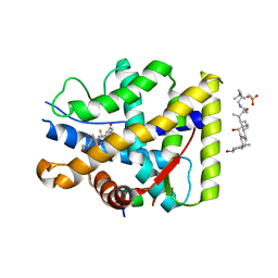
1ATP
 | | 2.2 angstrom refined crystal structure of the catalytic subunit of cAMP-dependent protein kinase complexed with MNATP and a peptide inhibitor | | 分子名称: | ADENOSINE-5'-TRIPHOSPHATE, MANGANESE (II) ION, PEPTIDE INHIBITOR PKI(5-24), ... | | 著者 | Zheng, J, Trafny, E.A, Knighton, D.R, Xuong, N.-H, Taylor, S.S, Teneyck, L.F, Sowadski, J.M. | | 登録日 | 1993-01-08 | | 公開日 | 1993-04-15 | | 最終更新日 | 2019-08-14 | | 実験手法 | X-RAY DIFFRACTION (2.2 Å) | | 主引用文献 | 2.2 A refined crystal structure of the catalytic subunit of cAMP-dependent protein kinase complexed with MnATP and a peptide inhibitor.
Acta Crystallogr.,Sect.D, 49, 1993
|
|
3NS9
 | | Crystal structure of CDK2 in complex with inhibitor BS-194 | | 分子名称: | (2S,3S)-3-{[7-(benzylamino)-3-(1-methylethyl)pyrazolo[1,5-a]pyrimidin-5-yl]amino}butane-1,2,4-triol, Cell division protein kinase 2 | | 著者 | Hazel, P, Freemont, P.S. | | 登録日 | 2010-07-01 | | 公開日 | 2010-12-08 | | 最終更新日 | 2023-09-06 | | 実験手法 | X-RAY DIFFRACTION (1.78 Å) | | 主引用文献 | A Novel Pyrazolo[1,5-a]pyrimidine Is a Potent Inhibitor of Cyclin-Dependent Protein Kinases 1, 2, and 9, Which Demonstrates Antitumor Effects in Human Tumor Xenografts Following Oral Administration.
J.Med.Chem., 53, 2010
|
|
1R2P
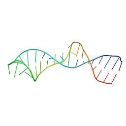 | | Solution structure of domain 5 from the ai5(gamma) group II intron | | 分子名称: | 34-MER | | 著者 | Sigel, R.K.O, Sashital, D.G, Abramovitz, D.L, Palmer III, A.G, Butcher, S.E, Pyle, A.M. | | 登録日 | 2003-09-29 | | 公開日 | 2004-02-03 | | 最終更新日 | 2024-05-22 | | 実験手法 | SOLUTION NMR | | 主引用文献 | Solution structure of domain 5 of a group II intron ribozyme reveals a new RNA motif.
Nat.Struct.Mol.Biol., 11, 2004
|
|
5I9E
 | |
1U9B
 | | MURINE/HUMAN UBIQUITIN-CONJUGATING ENZYME UBC9 | | 分子名称: | UBIQUITIN-CONJUGATING ENZYME E9 | | 著者 | Tong, H, Hateboer, G, Perrakis, A, Bernards, R, Sixma, T.K. | | 登録日 | 1997-05-20 | | 公開日 | 1997-07-07 | | 最終更新日 | 2024-05-22 | | 実験手法 | X-RAY DIFFRACTION (2 Å) | | 主引用文献 | Crystal structure of murine/human Ubc9 provides insight into the variability of the ubiquitin-conjugating system.
J.Biol.Chem., 272, 1997
|
|
6S1C
 | | P3221 crystal form of the Ctf18-1-8/Pol2(1-528) complex | | 分子名称: | Chromosome transmission fidelity protein 18, Chromosome transmission fidelity protein 8, DNA polymerase epsilon catalytic subunit A, ... | | 著者 | Grabarczyk, D.B. | | 登録日 | 2019-06-18 | | 公開日 | 2020-07-08 | | 最終更新日 | 2024-01-24 | | 実験手法 | X-RAY DIFFRACTION (6.1 Å) | | 主引用文献 | Ctf18-RFC and DNA Pol ε form a stable leading strand polymerase/clamp loader complex required for normal and perturbed DNA replication.
Nucleic Acids Res., 48, 2020
|
|
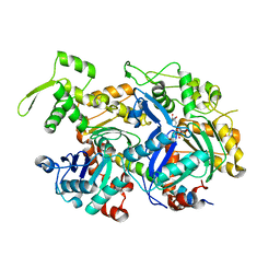
6YI9
 | | Crystal structure of the rat cytosolic PCK1, acetylated on Lys244 | | 分子名称: | 1,2-ETHANEDIOL, Phosphoenolpyruvate carboxykinase, cytosolic [GTP] | | 著者 | Latorre-Muro, P, Baeza, J, Hurtado-Guerrero, R, Hicks, T, Delso, I, Hernandez-Ruiz, C, Velazquez-Campoy, A, Lawton, A.J, Angulo, J, Denu, J.M, Carrodeguas, J.A. | | 登録日 | 2020-04-01 | | 公開日 | 2020-12-23 | | 最終更新日 | 2024-01-24 | | 実験手法 | X-RAY DIFFRACTION (1.75 Å) | | 主引用文献 | Self-acetylation at the active site of phosphoenolpyruvate carboxykinase (PCK1) controls enzyme activity.
J.Biol.Chem., 296, 2021
|
|
1U9A
 | | HUMAN UBIQUITIN-CONJUGATING ENZYME UBC9 | | 分子名称: | UBIQUITIN-CONJUGATING ENZYME | | 著者 | Tong, H, Hateboer, G, Perrakis, A, Bernards, R, Sixma, T.K. | | 登録日 | 1997-02-11 | | 公開日 | 1997-05-15 | | 最終更新日 | 2024-05-22 | | 実験手法 | X-RAY DIFFRACTION (2 Å) | | 主引用文献 | Crystal structure of murine/human Ubc9 provides insight into the variability of the ubiquitin-conjugating system.
J.Biol.Chem., 272, 1997
|
|
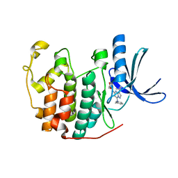
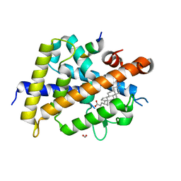
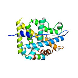
7C7V
 | | Vitamin D3 receptor/lithochoric acid derivative complex | | 分子名称: | (4R)-4-[(3R,5R,8R,9S,10S,13R,14S,17R)-10,13-dimethyl-3-(2-methyl-2-oxidanyl-propyl)-2,3,4,5,6,7,8,9,11,12,14,15,16,17-tetradecahydro-1H-cyclopenta[a]phenanthren-17-yl]pentanoic acid, FORMIC ACID, Mediator of RNA polymerase II transcription subunit 1, ... | | 著者 | Masuno, H, Numoto, N, Kagechika, H, Ito, N. | | 登録日 | 2020-05-26 | | 公開日 | 2021-01-20 | | 最終更新日 | 2023-11-29 | | 実験手法 | X-RAY DIFFRACTION (2 Å) | | 主引用文献 | Lithocholic Acid Derivatives as Potent Vitamin D Receptor Agonists.
J.Med.Chem., 64, 2021
|
|
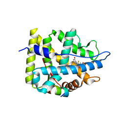
7C7W
 | | Vitamin D3 receptor/lithochoric acid derivative complex | | 分子名称: | (4R)-4-[(3S,5R,8R,9S,10S,13R,14S,17R)-10,13-dimethyl-3-(2-methyl-2-oxidanyl-propyl)-2,3,4,5,6,7,8,9,11,12,14,15,16,17-tetradecahydro-1H-cyclopenta[a]phenanthren-17-yl]pentanoic acid, FORMIC ACID, Mediator of RNA polymerase II transcription subunit 1, ... | | 著者 | Masuno, H, Numoto, N, Kagechika, H, Ito, N. | | 登録日 | 2020-05-26 | | 公開日 | 2021-01-20 | | 最終更新日 | 2023-11-29 | | 実験手法 | X-RAY DIFFRACTION (1.9 Å) | | 主引用文献 | Lithocholic Acid Derivatives as Potent Vitamin D Receptor Agonists.
J.Med.Chem., 64, 2021
|
|
6DGP
 | |
7YXP
 | | Crystal structure of WT AncGR2-LBD WT bound to dexamethasone and SHP coregulator fragment | | 分子名称: | 2-AMINO-2-HYDROXYMETHYL-PROPANE-1,3-DIOL, Ancestral Glucocorticoid Receptor2, DEXAMETHASONE, ... | | 著者 | Jimenez-Panizo, A, Estebanez-Perpina, E, Fuentes-Prior, P. | | 登録日 | 2022-02-16 | | 公開日 | 2022-12-07 | | 最終更新日 | 2024-01-31 | | 実験手法 | X-RAY DIFFRACTION (3.36 Å) | | 主引用文献 | The multivalency of the glucocorticoid receptor ligand-binding domain explains its manifold physiological activities.
Nucleic Acids Res., 50, 2022
|
|
7YXC
 | | Crystal structure of WT AncGR2-LBD bound to dexamethasone and SHP coregulator fragment | | 分子名称: | Ancestral Glucocorticoid Receptor2, CARBONATE ION, DEXAMETHASONE, ... | | 著者 | Jimenez-Panizo, A, Estebanez-Perpina, E, Fuentes-Prior, P. | | 登録日 | 2022-02-15 | | 公開日 | 2022-12-07 | | 最終更新日 | 2024-01-31 | | 実験手法 | X-RAY DIFFRACTION (2.25 Å) | | 主引用文献 | The multivalency of the glucocorticoid receptor ligand-binding domain explains its manifold physiological activities.
Nucleic Acids Res., 50, 2022
|
|
7YXR
 | | Crystal structure of mutant AncGR2-LBD (Y545A) bound to dexamethasone and SHP coregulator fragment | | 分子名称: | Ancestral Glucocorticoid Receptor2, DEXAMETHASONE, FORMIC ACID, ... | | 著者 | Jimenez-Panizo, A, Estebanez-Perpina, E, Fuentes-Prior, P. | | 登録日 | 2022-02-16 | | 公開日 | 2022-12-07 | | 最終更新日 | 2024-01-31 | | 実験手法 | X-RAY DIFFRACTION (2.5 Å) | | 主引用文献 | The multivalency of the glucocorticoid receptor ligand-binding domain explains its manifold physiological activities.
Nucleic Acids Res., 50, 2022
|
|
7YXD
 | | Crystal structure of WT AncGR2-LBD bound to dexamethasone and SHP coregulator fragment | | 分子名称: | Ancestral Glucocorticoid Receptor2, DEXAMETHASONE, SHP NR Box 1 Peptide, ... | | 著者 | Jimenez-Panizo, A, Estebanez-Perpina, E, Fuentes-Prior, P. | | 登録日 | 2022-02-15 | | 公開日 | 2022-12-07 | | 最終更新日 | 2024-01-31 | | 実験手法 | X-RAY DIFFRACTION (2.3 Å) | | 主引用文献 | The multivalency of the glucocorticoid receptor ligand-binding domain explains its manifold physiological activities.
Nucleic Acids Res., 50, 2022
|
|
7YXO
 | | Crystal structure of WT AncGR2-LBD bound to dexamethasone and SHP coregulator fragment | | 分子名称: | 3-[(3-CHOLAMIDOPROPYL)DIMETHYLAMMONIO]-1-PROPANESULFONATE, Ancestral Glucocorticoid Receptor2, DEXAMETHASONE, ... | | 著者 | Jimenez-Panizo, A, Estebanez-Perpina, E, Fuentes-Prior, P. | | 登録日 | 2022-02-16 | | 公開日 | 2022-12-07 | | 最終更新日 | 2024-01-31 | | 実験手法 | X-RAY DIFFRACTION (2.99 Å) | | 主引用文献 | The multivalency of the glucocorticoid receptor ligand-binding domain explains its manifold physiological activities.
Nucleic Acids Res., 50, 2022
|
|
7YXN
 | | Crystal structure of WT AncGR2-LBD bound to dexamethasone and SHP coregulator fragment | | 分子名称: | Ancestral Glucocorticoid Receptor2, DEXAMETHASONE, FORMIC ACID, ... | | 著者 | Jimenez-Panizo, A, Estebanez-Perpina, E, Fuentes-Prior, P. | | 登録日 | 2022-02-16 | | 公開日 | 2022-12-07 | | 最終更新日 | 2024-01-31 | | 実験手法 | X-RAY DIFFRACTION (2.46 Å) | | 主引用文献 | The multivalency of the glucocorticoid receptor ligand-binding domain explains its manifold physiological activities.
Nucleic Acids Res., 50, 2022
|
|
6GME
 | |
1B4L
 | | 15 ATMOSPHERE OXYGEN YEAST CU/ZN SUPEROXIDE DISMUTASE ROOM TEMPERATURE (298K) STRUCTURE | | 分子名称: | COPPER (II) ION, PROTEIN (CU/ZN SUPEROXIDE DISMUTASE), ZINC ION | | 著者 | Hart, P.J, Balbirnie, M.M, Ogihara, N.L, Nersissian, A.M, Weiss, M.S, Valentine, J.S, Eisenberg, D. | | 登録日 | 1998-12-22 | | 公開日 | 1999-12-23 | | 最終更新日 | 2023-12-27 | | 実験手法 | X-RAY DIFFRACTION (1.8 Å) | | 主引用文献 | A structure-based mechanism for copper-zinc superoxide dismutase.
Biochemistry, 38, 1999
|
|
3MY1
 | | Structure of CDK9/cyclinT1 in complex with DRB | | 分子名称: | 5,6-dichloro-1-beta-D-ribofuranosyl-1H-benzimidazole, Cell division protein kinase 9, Cyclin-T1, ... | | 著者 | Baumli, S, Johnson, L.N. | | 登録日 | 2010-05-09 | | 公開日 | 2010-09-29 | | 最終更新日 | 2023-11-01 | | 実験手法 | X-RAY DIFFRACTION (2.8 Å) | | 主引用文献 | Halogen bonds form the basis for selective P-TEFb inhibition by DRB
Chem.Biol., 17, 2010
|
|


