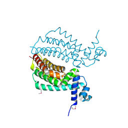
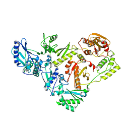
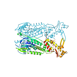
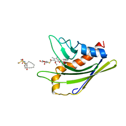
2IW5
 
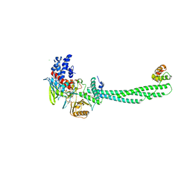 | | Structural Basis for CoREST-Dependent Demethylation of Nucleosomes by the Human LSD1 Histone Demethylase | | 分子名称: | AMMONIUM ION, CHLORIDE ION, FLAVIN-ADENINE DINUCLEOTIDE, ... | | 著者 | Yang, M, Gocke, C.B, Luo, X, Borek, D, Tomchick, D.R, Machius, M, Otwinowski, Z, Yu, H. | | 登録日 | 2006-06-26 | | 公開日 | 2006-08-09 | | 最終更新日 | 2024-05-08 | | 実験手法 | X-RAY DIFFRACTION (2.57 Å) | | 主引用文献 | Structural Basis for Corest-Dependent Demethylation of Nucleosomes by the Human Lsd1 Histone Demethylase
Mol.Cell, 23, 2006
|
|
3IH2
 | | TM1030 crystallized at 323K | | 分子名称: | Transcriptional regulator, TetR family | | 著者 | Koclega, K.D, Chruszcz, M, Bujacz, G, Joachimiak, A, Minor, W, Midwest Center for Structural Genomics (MCSG) | | 登録日 | 2009-07-29 | | 公開日 | 2009-08-11 | | 最終更新日 | 2023-11-22 | | 実験手法 | X-RAY DIFFRACTION (2.3 Å) | | 主引用文献 | 'Hot' macromolecular crystals.
Cryst.Growth Des., 10, 2010
|
|
3EZ2
 | | Partition protein-ADP complex | | 分子名称: | 4-(2-HYDROXYETHYL)-1-PIPERAZINE ETHANESULFONIC ACID, ADENOSINE-5'-DIPHOSPHATE, GLYCEROL, ... | | 著者 | Schumacher, M.A, Dunham, T.D, Xu, W, Funnell, B. | | 登録日 | 2008-10-22 | | 公開日 | 2009-06-02 | | 最終更新日 | 2023-09-06 | | 実験手法 | X-RAY DIFFRACTION (2.05 Å) | | 主引用文献 | Structural basis for ADP-mediated transcriptional regulation by P1 and P7 ParA.
Embo J., 28, 2009
|
|
3VW2
 | | Crystal Strucuture of The Berberine-bound Form of RamR (Transcriptional Regurator of TetR Family) from Salmonella Typhimurium | | 分子名称: | BERBERINE, Putative regulatory protein, SULFATE ION | | 著者 | Sakurai, K, Nikaido, E, Nakashima, R, Yamasaki, S, Yamaguchi, A, Nishino, K. | | 登録日 | 2012-07-30 | | 公開日 | 2013-07-03 | | 最終更新日 | 2024-03-20 | | 実験手法 | X-RAY DIFFRACTION (2.34 Å) | | 主引用文献 | The crystal structure of multidrug-resistance regulator RamR with multiple drugs
Nat Commun, 4, 2013
|
|
3IH4
 | | TM1030 crystallized at 277K | | 分子名称: | Transcriptional regulator, TetR family | | 著者 | Koclega, K.D, Chruszcz, M, Bujacz, G, Joachimiak, A, Minor, W, Midwest Center for Structural Genomics (MCSG) | | 登録日 | 2009-07-29 | | 公開日 | 2009-08-11 | | 最終更新日 | 2023-11-22 | | 実験手法 | X-RAY DIFFRACTION (2.3 Å) | | 主引用文献 | 'Hot' macromolecular crystals.
Cryst.Growth Des., 10, 2010
|
|
1KCA
 | |
6N2I
 | | Lon protease AAA+ domain | | 分子名称: | ADENOSINE-5'-DIPHOSPHATE, DNA-binding ATP-dependent protease La | | 著者 | Botos, I, Li, M, Wlodawer, A, Gustchina, A. | | 登録日 | 2018-11-13 | | 公開日 | 2019-07-10 | | 最終更新日 | 2023-10-11 | | 実験手法 | X-RAY DIFFRACTION (3.5 Å) | | 主引用文献 | New insights into structural and functional relationships between LonA proteases and ClpB chaperones.
Febs Open Bio, 9, 2019
|
|
3EZ6
 | | Structure of parA-ADP complex:tetragonal form | | 分子名称: | ADENOSINE-5'-DIPHOSPHATE, MAGNESIUM ION, Plasmid partition protein A | | 著者 | Schumacher, M.A. | | 登録日 | 2008-10-22 | | 公開日 | 2009-06-02 | | 最終更新日 | 2023-09-06 | | 実験手法 | X-RAY DIFFRACTION (2.58 Å) | | 主引用文献 | Structural basis for ADP-mediated transcriptional regulation by P1 and P7 ParA.
Embo J., 28, 2009
|
|
3IH3
 | | TM1030 crystallized at 310K | | 分子名称: | Transcriptional regulator, TetR family | | 著者 | Koclega, K.D, Chruszcz, M, Bujacz, G, Joachimiak, A, Minor, W, Midwest Center for Structural Genomics (MCSG) | | 登録日 | 2009-07-29 | | 公開日 | 2009-08-11 | | 最終更新日 | 2023-11-22 | | 実験手法 | X-RAY DIFFRACTION (2.35 Å) | | 主引用文献 | 'Hot' macromolecular crystals.
Cryst.Growth Des., 10, 2010
|
|
3IG1
 | | HIV-1 Reverse Transcriptase with the Inhibitor beta-Thujaplicinol Bound at the RNase H Active Site | | 分子名称: | 2,7-dihydroxy-4-(propan-2-yl)cyclohepta-2,4,6-trien-1-one, HIV-1 Reverse Transcriptase p51 subunit, HIV-1 Reverse Transcriptase p66 subunit, ... | | 著者 | Himmel, D.M, Maegley, K.A, Pauly, T.A, Arnold, E. | | 登録日 | 2009-07-27 | | 公開日 | 2010-01-26 | | 最終更新日 | 2023-09-06 | | 実験手法 | X-RAY DIFFRACTION (2.8 Å) | | 主引用文献 | Structure of HIV-1 reverse transcriptase with the inhibitor beta-Thujaplicinol bound at the RNase H active site.
Structure, 17, 2009
|
|
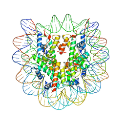
5UJM
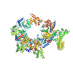 | | Structure of the active form of human Origin Recognition Complex and its ATPase motor module | | 分子名称: | ADENOSINE-5'-TRIPHOSPHATE, MAGNESIUM ION, Origin recognition complex subunit 1, ... | | 著者 | Tocilj, A, On, K, Yuan, Z, Sun, J, Elkayam, E, Li, H, Stillman, B, Joshua-Tor, L. | | 登録日 | 2017-01-18 | | 公開日 | 2017-02-08 | | 最終更新日 | 2024-03-06 | | 実験手法 | ELECTRON MICROSCOPY (18 Å) | | 主引用文献 | Structure of the active form of human Origin Recognition Complex and its ATPase motor module.
Elife, 6, 2017
|
|
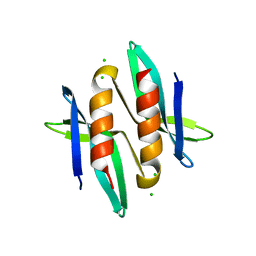
4DRA
 | | Crystal structure of MHF complex | | 分子名称: | Centromere protein S, Centromere protein X | | 著者 | Tao, Y, Niu, L, Teng, M. | | 登録日 | 2012-02-17 | | 公開日 | 2012-05-16 | | 最終更新日 | 2024-03-20 | | 実験手法 | X-RAY DIFFRACTION (2.414 Å) | | 主引用文献 | The structure of the FANCM-MHF complex reveals physical features for functional assembly
Nat Commun, 3, 2012
|
|
5A4N
 | |
4Y17
 | | SdiA in complex with 3-oxo-C8-homoserine lactone | | 分子名称: | 3-OXO-OCTANOIC ACID (2-OXO-TETRAHYDRO-FURAN-3-YL)-AMIDE, Transcriptional regulator of ftsQAZ gene cluster | | 著者 | Nguyen, N.X, Nguyen, Y, Sperandio, V, Jiang, Y. | | 登録日 | 2015-02-06 | | 公開日 | 2015-04-08 | | 最終更新日 | 2023-09-27 | | 実験手法 | X-RAY DIFFRACTION (2.84 Å) | | 主引用文献 | Structural and Mechanistic Roles of Novel Chemical Ligands on the SdiA Quorum-Sensing Transcription Regulator.
Mbio, 6, 2015
|
|
1AYZ
 | |
6RWO
 | | SIVrcm intasome (Q148H/G140S) in complex with bictegravir | | 分子名称: | Bictegravir, CHLORIDE ION, DNA (5'-D(*AP*AP*CP*TP*GP*GP*TP*AP*GP*AP*GP*AP*TP*TP*TP*TP*TP*CP*TP*TP*AP*GP*C)-3'), ... | | 著者 | Cherepanov, P, Nans, A, Cook, N. | | 登録日 | 2019-06-05 | | 公開日 | 2020-02-05 | | 最終更新日 | 2024-07-10 | | 実験手法 | ELECTRON MICROSCOPY (3.05 Å) | | 主引用文献 | Structural basis of second-generation HIV integrase inhibitor action and viral resistance.
Science, 367, 2020
|
|
3SXY
 | |
3P5J
 | |
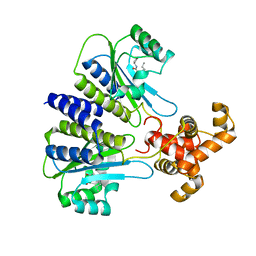
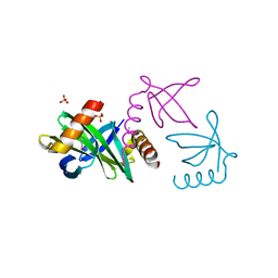
4EOG
 | | Crystal structure of Csx1 of Pyrococcus furiosus | | 分子名称: | Putative uncharacterized protein, SULFATE ION, ZINC ION | | 著者 | Kim, Y.K, Oh, B.H. | | 登録日 | 2012-04-14 | | 公開日 | 2013-01-02 | | 最終更新日 | 2013-07-24 | | 実験手法 | X-RAY DIFFRACTION (2.3 Å) | | 主引用文献 | Crystal structure and nucleic acid-binding activity of the CRISPR-associated protein Csx1 of Pyrococcus furiosus.
Proteins, 81, 2013
|
|
6JUI
 | | The atypical Myb-like protein Cdc5 contains two distinct nucleic acid-binding surfaces | | 分子名称: | Pre-mRNA-splicing factor CEF1 | | 著者 | Wang, C, Li, G, Li, M, Yang, J, Liu, J. | | 登録日 | 2019-04-14 | | 公開日 | 2020-02-19 | | 最終更新日 | 2024-03-27 | | 実験手法 | X-RAY DIFFRACTION (2.402 Å) | | 主引用文献 | Two distinct nucleic acid binding surfaces of Cdc5 regulate development.
Biochem.J., 476, 2019
|
|
6K5U
 | |
2XIW
 | | Crystal structure of the Sac7d-derived IgG1-binder C3-C24S | | 分子名称: | CHLORIDE ION, DNA-BINDING PROTEIN 7D, SULFATE ION | | 著者 | Bellinzoni, M, Colinet, S, Behar, G, Alzari, P.M, Pecorari, F. | | 登録日 | 2010-07-01 | | 公開日 | 2011-07-13 | | 最終更新日 | 2013-04-10 | | 実験手法 | X-RAY DIFFRACTION (1.5 Å) | | 主引用文献 | Tolerance of the Archaeal Sac7D Scaffold Protein to Alternative Library Designs: Characterization of Anti-Immunoglobulin G Affitins.
Protein Eng.Des.Sel., 26, 2013
|
|
2NQB
 | |
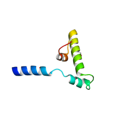
2L65
 | | HADDOCK calculated model of the complex of the resistance protein CalC and Calicheamicin-Gamma | | 分子名称: | 2,4-dideoxy-4-(ethylamino)-3-O-methyl-alpha-L-threo-pentopyranose-(1-2)-4-amino-4,6-dideoxy-beta-D-glucopyranose, 2,6-dideoxy-4-thio-beta-D-allopyranose, 3-O-methyl-alpha-L-rhamnopyranose, ... | | 著者 | Singh, S, Markley, J.L, Thorson, J.S, Center for Eukaryotic Structural Genomics (CESG) | | 登録日 | 2010-11-15 | | 公開日 | 2011-03-02 | | 最終更新日 | 2024-05-01 | | 実験手法 | SOLUTION NMR | | 主引用文献 | Structural insight into the self-sacrifice mechanism of enediyne resistance.
Acs Chem.Biol., 1, 2006
|
|
8BUO
 | | Structure of DDB1 bound to DS24-engaged CDK12-cyclin K | | 分子名称: | (2~{R})-2-[[6-[(3-fluoranyl-4-pyridin-2-yl-phenyl)methylamino]-9-propan-2-yl-purin-2-yl]amino]butan-1-ol, Cyclin-K, Cyclin-dependent kinase 12, ... | | 著者 | Kozicka, Z, Kempf, G, Petzold, G, Thoma, N.H. | | 登録日 | 2022-11-30 | | 公開日 | 2023-09-13 | | 最終更新日 | 2024-01-03 | | 実験手法 | X-RAY DIFFRACTION (3.58 Å) | | 主引用文献 | Design principles for cyclin K molecular glue degraders.
Nat.Chem.Biol., 20, 2024
|
|


