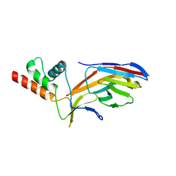
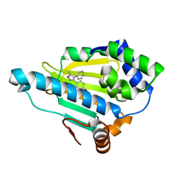
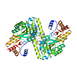
5TYA
 
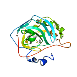 | | Identification of a New Zinc Binding Chemotype by Fragment Screening | | 分子名称: | (5R)-5-phenyl-1,3-thiazolidine-2,4-dione, Carbonic anhydrase 2, ZINC ION | | 著者 | Peat, T.S, Poulsen, S.A, Ren, B, Dolezal, O, Woods, L.A, Mujumdar, P, Chrysanthopoulos, P.K. | | 登録日 | 2016-11-18 | | 公開日 | 2017-08-30 | | 最終更新日 | 2023-10-04 | | 実験手法 | X-RAY DIFFRACTION (1.5 Å) | | 主引用文献 | Identification of a New Zinc Binding Chemotype by Fragment Screening.
J. Med. Chem., 60, 2017
|
|
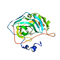
6FKZ
 | |
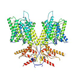
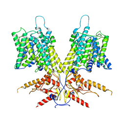
7S9E
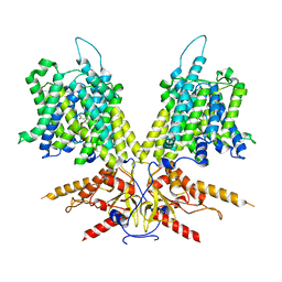 | | Cryo-EM Structure of dolphin Prestin: Inhibited II (Sulfate +Salicylate) state | | 分子名称: | 2-HYDROXYBENZOIC ACID, Prestin | | 著者 | Bavi, N, Clark, M.D, Contreras, G.F, Shen, R, Reddy, B.G, Milewski, W, Perozo, E. | | 登録日 | 2021-09-20 | | 公開日 | 2021-11-03 | | 最終更新日 | 2024-06-05 | | 実験手法 | ELECTRON MICROSCOPY (3.7 Å) | | 主引用文献 | The conformational cycle of prestin underlies outer-hair cell electromotility.
Nature, 600, 2021
|
|
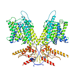
7S8X
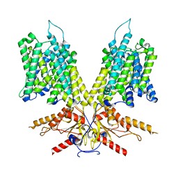 | | Cryo-EM Structure of dolphin Prestin: Sensor Up (compact) state | | 分子名称: | Prestin | | 著者 | Bavi, N, Clark, M.D, Contreras, G.F, Shen, R, Reddy, B.G, Milewski, W, Perozo, E. | | 登録日 | 2021-09-20 | | 公開日 | 2021-11-03 | | 最終更新日 | 2024-06-05 | | 実験手法 | ELECTRON MICROSCOPY (3.3 Å) | | 主引用文献 | The conformational cycle of prestin underlies outer-hair cell electromotility.
Nature, 600, 2021
|
|
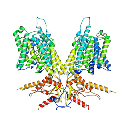
7S9C
 | | Cryo-EM Structure of dolphin Prestin: Sensor Down II (Expanded II) state | | 分子名称: | Prestin | | 著者 | Bavi, N, Clark, M.D, Contreras, G.F, Shen, R, Reddy, B.G, Milewski, W, Perozo, E. | | 登録日 | 2021-09-20 | | 公開日 | 2021-11-03 | | 最終更新日 | 2024-06-05 | | 実験手法 | ELECTRON MICROSCOPY (6.7 Å) | | 主引用文献 | The conformational cycle of prestin underlies outer-hair cell electromotility.
Nature, 600, 2021
|
|
7S9D
 | | Cryo-EM Structure of dolphin Prestin: Intermediate state | | 分子名称: | Prestin | | 著者 | Bavi, N, Clark, M.D, Contreras, G.F, Shen, R, Reddy, B.G, Milewski, W, Perozo, E. | | 登録日 | 2021-09-20 | | 公開日 | 2021-11-03 | | 最終更新日 | 2024-06-05 | | 実験手法 | ELECTRON MICROSCOPY (4.6 Å) | | 主引用文献 | The conformational cycle of prestin underlies outer-hair cell electromotility.
Nature, 600, 2021
|
|
7O3B
 | |
7S9A
 | | Cryo-EM Structure of dolphin Prestin: Inhibited I (Chloride + Salicylate) | | 分子名称: | 2-HYDROXYBENZOIC ACID, Prestin | | 著者 | Bavi, N, Clark, M.D, Contreras, G.F, Shen, R, Reddy, B.G, Milewski, W, Perozo, E. | | 登録日 | 2021-09-20 | | 公開日 | 2021-11-03 | | 最終更新日 | 2024-06-05 | | 実験手法 | ELECTRON MICROSCOPY (3.8 Å) | | 主引用文献 | The conformational cycle of prestin underlies outer-hair cell electromotility.
Nature, 600, 2021
|
|
7O0S
 | |
7S9B
 | | Cryo-EM Structure of dolphin Prestin: Sensor Down I (Expanded) state | | 分子名称: | Prestin | | 著者 | Bavi, N, Clark, M.D, Contreras, G.F, Shen, R, Reddy, B.G, Milewski, W, Perozo, E. | | 登録日 | 2021-09-20 | | 公開日 | 2021-11-03 | | 最終更新日 | 2024-06-05 | | 実験手法 | ELECTRON MICROSCOPY (4.2 Å) | | 主引用文献 | The conformational cycle of prestin underlies outer-hair cell electromotility.
Nature, 600, 2021
|
|
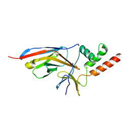
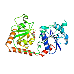
4HS2
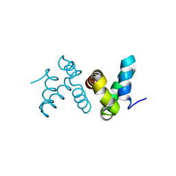 | | Crystal Structure of the Human SPOP C-terminal Domain | | 分子名称: | Speckle-type POZ protein | | 著者 | Van Geersdaele, L.K, Stead, M.A, Carr, S.B, Wright, S.C. | | 登録日 | 2012-10-29 | | 公開日 | 2013-09-11 | | 最終更新日 | 2024-02-28 | | 実験手法 | X-RAY DIFFRACTION (1.53 Å) | | 主引用文献 | Structural basis of high-order oligomerization of the cullin-3 adaptor SPOP.
Acta Crystallogr.,Sect.D, 69, 2013
|
|
6CB1
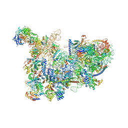 | | Yeast nucleolar pre-60S ribosomal subunit (state 3) | | 分子名称: | 35S pre-ribosomal RNA miscRNA, 5.8S rRNA, 60S ribosomal protein L13-A, ... | | 著者 | Sanghai, Z.A, Miller, L, Barandun, J, Hunziker, M, Chaker-Margot, M, Klinge, S. | | 登録日 | 2018-02-01 | | 公開日 | 2018-03-14 | | 最終更新日 | 2024-03-13 | | 実験手法 | ELECTRON MICROSCOPY (4.6 Å) | | 主引用文献 | Modular assembly of the nucleolar pre-60S ribosomal subunit.
Nature, 556, 2018
|
|
2YJW
 | | Tricyclic series of Hsp90 inhibitors | | 分子名称: | 4-(5-METHYL-4-PHENYLISOXAZOL-3-YL)BENZENE-1,3-DIOL, HEAT SHOCK PROTEIN HSP 90-ALPHA | | 著者 | Dupuy, A, Vallee, F. | | 登録日 | 2011-05-24 | | 公開日 | 2011-10-19 | | 最終更新日 | 2024-05-08 | | 実験手法 | X-RAY DIFFRACTION (1.61 Å) | | 主引用文献 | Tricyclic Series of Heat Shock Protein 90 (Hsp90) Inhibitors Part I: Discovery of Tricyclic Imidazo[4,5-C]Pyridines as Potent Inhibitors of the Hsp90 Molecular Chaperone.
J.Med.Chem., 54, 2011
|
|
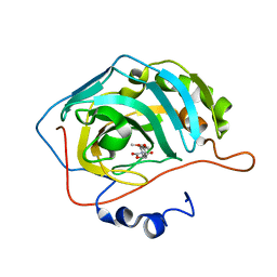
5U0F
 | | Identification of a New Zinc Binding Chemotype by Fragment Screening | | 分子名称: | (5R)-5-[(2,4-dimethoxyphenyl)methyl]-2-sulfanylidene-1,3-thiazolidin-4-one, Carbonic anhydrase 2, ZINC ION | | 著者 | Peat, T.S, Poulsen, S.A, Ren, B, Dolezal, O, Woods, L.A, Mujumdar, P, Chrysanthopoulos, P.K. | | 登録日 | 2016-11-23 | | 公開日 | 2017-08-30 | | 最終更新日 | 2023-10-04 | | 実験手法 | X-RAY DIFFRACTION (1.21 Å) | | 主引用文献 | Identification of a New Zinc Binding Chemotype by Fragment Screening.
J. Med. Chem., 60, 2017
|
|
1NNU
 | | Crystal Structure Analysis of Plasmodium falciparum enoyl-acyl-carrier-protein reductase with Triclosan Analog | | 分子名称: | 6-(4-CHLORO-2-HYDROXY-PHENOXY)-NAPHTHALEN-2-OL, NICOTINAMIDE-ADENINE-DINUCLEOTIDE, enoyl-acyl carrier reductase | | 著者 | Perozzo, R, Kuo, M, Sidhu, A.S, Valiyaveettil, J.T, Bittman, R, Jacobs Jr, W.R, Fidock, D.A, Sacchettini, J.C. | | 登録日 | 2003-01-14 | | 公開日 | 2003-02-25 | | 最終更新日 | 2024-02-14 | | 実験手法 | X-RAY DIFFRACTION (2.5 Å) | | 主引用文献 | Structural Elucidation of the Specificity of the Antibacterial Agent Triclosan for
Malarial Enoyl Acyl Carrier Protein Reductase
J.Biol.Chem., 277, 2002
|
|
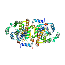
5TY9
 | | Identification of a New Zinc Binding Chemotype by Fragment Screening | | 分子名称: | (5R)-5-(2,4-dimethoxyphenyl)-1,3-oxazolidine-2,4-dione, Carbonic anhydrase 2, ZINC ION | | 著者 | Peat, T.S, Poulsen, S.A, Ren, B, Dolezal, O, Woods, L.A, Mujumdar, P, Chrysanthopoulos, P.K. | | 登録日 | 2016-11-18 | | 公開日 | 2017-08-30 | | 最終更新日 | 2023-10-04 | | 実験手法 | X-RAY DIFFRACTION (1.53 Å) | | 主引用文献 | Identification of a New Zinc Binding Chemotype by Fragment Screening.
J. Med. Chem., 60, 2017
|
|
4EU1
 | |
2BGU
 | | CRYSTAL STRUCTURE OF THE DNA MODIFYING ENZYME BETA-GLUCOSYLTRANSFERASE IN THE PRESENCE AND ABSENCE OF THE SUBSTRATE URIDINE DIPHOSPHOGLUCOSE | | 分子名称: | BETA-GLUCOSYLTRANSFERASE, URIDINE-5'-DIPHOSPHATE | | 著者 | Vrielink, A, Rueger, W, Driessen, H.P.C, Freemont, P.S. | | 登録日 | 1994-06-09 | | 公開日 | 1995-12-09 | | 最終更新日 | 2024-02-14 | | 実験手法 | X-RAY DIFFRACTION (2.2 Å) | | 主引用文献 | Crystal structure of the DNA modifying enzyme beta-glucosyltransferase in the presence and absence of the substrate uridine diphosphoglucose.
EMBO J., 13, 1994
|
|
3OJ2
 | |
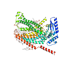
1OAA
 | | MOUSE SEPIAPTERIN REDUCTASE COMPLEXED WITH NADP AND OXALOACETATE | | 分子名称: | NADP NICOTINAMIDE-ADENINE-DINUCLEOTIDE PHOSPHATE, OXALOACETATE ION, SEPIAPTERIN REDUCTASE, ... | | 著者 | Auerbach, G, Herrmann, A, Bacher, A, Huber, R. | | 登録日 | 1997-08-25 | | 公開日 | 1999-02-16 | | 最終更新日 | 2024-02-14 | | 実験手法 | X-RAY DIFFRACTION (1.25 Å) | | 主引用文献 | The 1.25 A crystal structure of sepiapterin reductase reveals its binding mode to pterins and brain neurotransmitters.
EMBO J., 16, 1997
|
|
7RXH
 | |
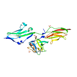
2BTC
 | | BOVINE TRYPSIN IN COMPLEX WITH SQUASH SEED INHIBITOR (CUCURBITA PEPO TRYPSIN INHIBITOR II) | | 分子名称: | CALCIUM ION, PROTEIN (TRYPSIN INHIBITOR), PROTEIN (TRYPSIN) | | 著者 | Helland, R, Berglund, G.I, Otlewski, J, Apostoluk, W, Andersen, O.A, Willassen, N.P, Smalas, A.O. | | 登録日 | 1998-12-11 | | 公開日 | 2000-01-19 | | 最終更新日 | 2023-08-23 | | 実験手法 | X-RAY DIFFRACTION (1.5 Å) | | 主引用文献 | High-resolution structures of three new trypsin-squash-inhibitor complexes: a detailed comparison with other trypsins and their complexes.
Acta Crystallogr.,Sect.D, 55, 1999
|
|
1NT9
 | | Complete 12-subunit RNA polymerase II | | 分子名称: | DNA-DIRECTED RNA POLYMERASE II 13.6 KD POLYPEPTIDE, DNA-DIRECTED RNA POLYMERASE II 19 KD POLYPEPTIDE, DNA-DIRECTED RNA POLYMERASE II LARGEST SUBUNIT, ... | | 著者 | Armache, K.-J, Kettenberger, H, Cramer, P. | | 登録日 | 2003-01-29 | | 公開日 | 2003-04-22 | | 最終更新日 | 2023-08-16 | | 実験手法 | X-RAY DIFFRACTION (4.2 Å) | | 主引用文献 | Architecture of initiation-competent 12-subunit RNA polymerase II
Proc.Natl.Acad.Sci.USA, 100, 2003
|
|
8CNK
 | |
6CHA
 | |


