6UWI
 
 | |
8A6I
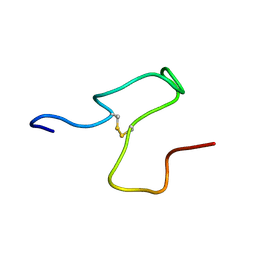
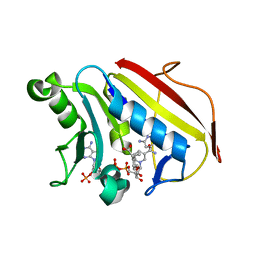
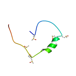 | | Structure of the low complexity domain of TDP-43 (fragment 309-350) with methionine sulfoxide modifications | | 分子名称: | TAR DNA-binding protein 43 | | 著者 | Carrasco, J, Anton, R, Pantoja-Uceda, D, Laurents, D.V, Oroz, J. | | 登録日 | 2022-06-17 | | 公開日 | 2023-02-08 | | 実験手法 | SOLUTION NMR | | 主引用文献 | Metamorphism in TDP-43 prion-like domain determines chaperone recognition.
Nat Commun, 14, 2023
|
|
3NWQ
 | |
8Q6U
 | |
3NY1
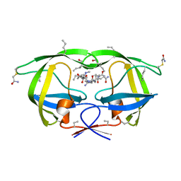
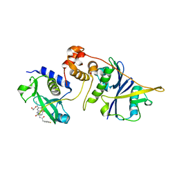
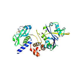
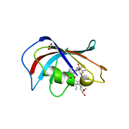
 | | Structure of the ubr-box of the UBR1 ubiquitin ligase | | 分子名称: | E3 ubiquitin-protein ligase UBR1, ZINC ION | | 著者 | Matta-Camacho, E, Kozlov, G, Li, F, Gehring, K. | | 登録日 | 2010-07-14 | | 公開日 | 2010-08-11 | | 最終更新日 | 2024-02-21 | | 実験手法 | X-RAY DIFFRACTION (2.085 Å) | | 主引用文献 | Structural basis of substrate recognition and specificity in the N-end rule pathway.
Nat.Struct.Mol.Biol., 17, 2010
|
|
4HNJ
 | |
3NZZ
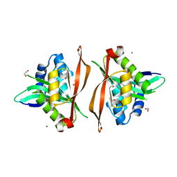
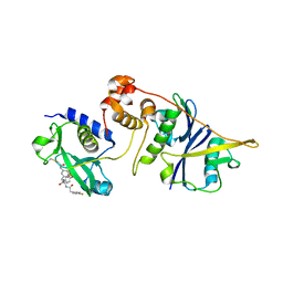
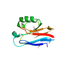
 | | Crystal Structure of the Salmonella Type III Secretion System Tip Protein SipD | | 分子名称: | Cell invasion protein sipD, NICKEL (II) ION | | 著者 | Chatterjee, S, Zhong, D, Nordhues, B.A, Battaile, K.P, Lovell, S, DeGuzman, R.N. | | 登録日 | 2010-07-18 | | 公開日 | 2010-11-17 | | 最終更新日 | 2024-02-21 | | 実験手法 | X-RAY DIFFRACTION (1.65 Å) | | 主引用文献 | The crystal structures of the Salmonella type III secretion system tip protein SipD in complex with deoxycholate and chenodeoxycholate.
Protein Sci., 20, 2011
|
|
4HP4
 | |
6UZ4
 | |
4HQA
 | |
7ZLO
 | | Crystal structure of SOCS2:ElonginB:ElonginC in complex with compound 12 | | 分子名称: | Elongin-B, Elongin-C, Suppressor of cytokine signaling 2, ... | | 著者 | Ramachandran, S, Ciulli, A, Makukhin, N. | | 登録日 | 2022-04-15 | | 公開日 | 2023-04-26 | | 最終更新日 | 2024-05-01 | | 実験手法 | X-RAY DIFFRACTION (2.22 Å) | | 主引用文献 | Structure-based design of a phosphotyrosine-masked covalent ligand targeting the E3 ligase SOCS2.
Nat Commun, 14, 2023
|
|
7ZLP
 | | Crystal structure of SOCS2:ElonginB:ElonginC in complex with compound 9 | | 分子名称: | Elongin-B, Elongin-C, PHOSPHATE ION, ... | | 著者 | Ramachandran, S, Ciulli, A, Makukhin, N. | | 登録日 | 2022-04-15 | | 公開日 | 2023-04-26 | | 最終更新日 | 2024-05-01 | | 実験手法 | X-RAY DIFFRACTION (1.94 Å) | | 主引用文献 | Structure-based design of a phosphotyrosine-masked covalent ligand targeting the E3 ligase SOCS2.
Nat Commun, 14, 2023
|
|
7ZLN
 | | Crystal structure of SOCS2:ElonginB:ElonginC in complex with compound 11 | | 分子名称: | Elongin-B, Elongin-C, Suppressor of cytokine signaling 2, ... | | 著者 | Ramachandran, S, Ciulli, A, Makukhin, N. | | 登録日 | 2022-04-15 | | 公開日 | 2023-04-26 | | 最終更新日 | 2024-05-01 | | 実験手法 | X-RAY DIFFRACTION (2.6 Å) | | 主引用文献 | Structure-based design of a phosphotyrosine-masked covalent ligand targeting the E3 ligase SOCS2.
Nat Commun, 14, 2023
|
|
7ZLS
 | | co-crystal structure of SOCS2:ElonginB:ElonginC in complex with compound 13 | | 分子名称: | 1,2-ETHANEDIOL, Elongin-B, Elongin-C, ... | | 著者 | Ramachandran, S, Ciulli, A, Makukhin, N. | | 登録日 | 2022-04-15 | | 公開日 | 2023-04-26 | | 最終更新日 | 2024-05-01 | | 実験手法 | X-RAY DIFFRACTION (1.92 Å) | | 主引用文献 | Structure-based design of a phosphotyrosine-masked covalent ligand targeting the E3 ligase SOCS2.
Nat Commun, 14, 2023
|
|
7ZLR
 | | Crystal structure of SOCS2:ElonginB:ElonginC in complex with compound 13 | | 分子名称: | Elongin-B, Elongin-C, Suppressor of cytokine signaling 2, ... | | 著者 | Ramachandran, S, Ciulli, A, Makukhin, N. | | 登録日 | 2022-04-15 | | 公開日 | 2023-04-26 | | 最終更新日 | 2024-05-01 | | 実験手法 | X-RAY DIFFRACTION (2.01 Å) | | 主引用文献 | Structure-based design of a phosphotyrosine-masked covalent ligand targeting the E3 ligase SOCS2.
Nat Commun, 14, 2023
|
|
7ZLM
 | | Crystal structure of SOCS2:ElonginB:ElonginC in complex with compound MN551 | | 分子名称: | Elongin-B, Elongin-C, Suppressor of cytokine signaling 2, ... | | 著者 | Ramachandran, S, Ciulli, A, Makukhin, N. | | 登録日 | 2022-04-15 | | 公開日 | 2023-04-26 | | 最終更新日 | 2024-05-01 | | 実験手法 | X-RAY DIFFRACTION (1.79 Å) | | 主引用文献 | Structure-based design of a phosphotyrosine-masked covalent ligand targeting the E3 ligase SOCS2.
Nat Commun, 14, 2023
|
|
6UWT
 | |
8S9Y
 | |
8A0W
 | |
7ZMX
 | |
6V9K
 | |
4HRS
 | |
6VCJ
 | | Crystal structure of hsDHFR in complex with NADP+, DAP, and R-naproxen | | 分子名称: | (2R)-2-(6-methoxynaphthalen-2-yl)propanoic acid, Dihydrofolate reductase, FOLIC ACID, ... | | 著者 | Pedersen, L.C, London, R.E, Gabel, S.A, Krahn, J.M, DeRose, E.F. | | 登録日 | 2019-12-21 | | 公開日 | 2020-10-28 | | 最終更新日 | 2023-10-11 | | 実験手法 | X-RAY DIFFRACTION (2.34 Å) | | 主引用文献 | The Structural Basis for Nonsteroidal Anti-Inflammatory Drug Inhibition of Human Dihydrofolate Reductase.
J.Med.Chem., 63, 2020
|
|
6VCU
 | | Homo sapiens FKBP12 protein bound with APX879 in P32 space group | | 分子名称: | ACETATE ION, N'-[(3S,4R,5S,8R,9E,12S,14S,15R,16S,18R,19R,26aS)-5,19-dihydroxy-3-{(1E)-1-[(1R,3R,4R)-4-hydroxy-3-methoxycyclohexyl]prop-1-en-2-yl}-14,16-dimethoxy-4,10,12,18-tetramethyl-1,20,21-trioxo-8-(prop-2-en-1-yl)-1,3,4,5,6,8,11,12,13,14,15,16,17,18,19,20,21,23,24,25,26,26a-docosahydro-7H-15,19-epoxypyrido[2,1-c][1,4]oxazacyclotricosin-7-ylidene]acetohydrazide, Peptidyl-prolyl cis-trans isomerase FKBP1A | | 著者 | Gobeil, S, Spicer, L. | | 登録日 | 2019-12-23 | | 公開日 | 2020-12-16 | | 最終更新日 | 2023-10-11 | | 実験手法 | X-RAY DIFFRACTION (1.69 Å) | | 主引用文献 | Leveraging Fungal and Human Calcineurin-Inhibitor Structures, Biophysical Data, and Dynamics To Design Selective and Nonimmunosuppressive FK506 Analogs.
Mbio, 12, 2021
|
|
4HZ1
 | | Crystal Structure of Pseudomonas aeruginosa azurin with iron(II) at the copper-binding site. | | 分子名称: | ACETATE ION, Azurin, FE (II) ION | | 著者 | McLaughlin, M.P, Retegan, M, Bill, E, Payne, T.M, Shafaat, H.S, Pea, S, Sudhamsu, J, Ensign, A.A, Crane, B.R, Neese, F, Holland, P.L. | | 登録日 | 2012-11-14 | | 公開日 | 2012-12-12 | | 最終更新日 | 2023-09-20 | | 実験手法 | X-RAY DIFFRACTION (2.2 Å) | | 主引用文献 | Azurin as a Protein Scaffold for a Low-coordinate Nonheme Iron Site with a Small-molecule Binding Pocket.
J.Am.Chem.Soc., 134, 2012
|
|


