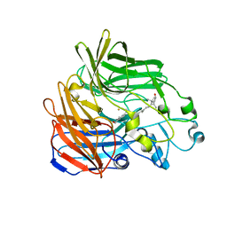
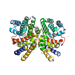
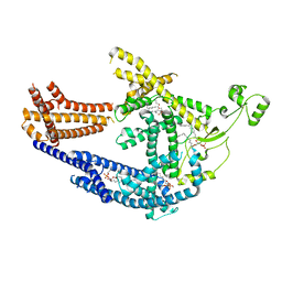
2CBO
 
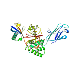
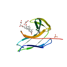 | | Crystal structure of the neocarzinostatin 3Tes24 mutant bound to testosterone hemisuccinate. | | 分子名称: | NEOCARZINOSTATIN, SULFATE ION, TESTOSTERONE HEMISUCCINATE | | 著者 | Drevelle, A, Graille, M, Heyd, B, Sorel, I, Ulryck, N, Pecorari, F, Desmadril, M, van Tilbeurgh, H, Minard, P. | | 登録日 | 2006-01-06 | | 公開日 | 2006-03-22 | | 最終更新日 | 2023-12-13 | | 実験手法 | X-RAY DIFFRACTION (1.7 Å) | | 主引用文献 | Structures of in Vitro Evolved Binding Sites on Neocarzinostatin Scaffold Reveal Unanticipated Evolutionary Pathways.
J.Mol.Biol., 358, 2006
|
|
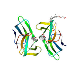
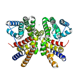
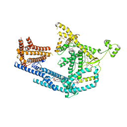
2CBT
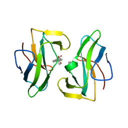 | | Crystal structure of the neocarzinostatin 4Tes1 mutant bound testosterone hemisuccinate. | | 分子名称: | NEOCARZINOSTATIN, TESTOSTERONE HEMISUCCINATE | | 著者 | Drevelle, A, Graille, M, Heyd, B, Sorel, I, Ulryck, N, Pecorari, F, Desmadril, M, Van Tilbeurgh, H, Minard, P. | | 登録日 | 2006-01-06 | | 公開日 | 2006-03-22 | | 最終更新日 | 2023-12-13 | | 実験手法 | X-RAY DIFFRACTION (2.2 Å) | | 主引用文献 | Structures of in Vitro Evolved Binding Sites on Neocarzinostatin Scaffold Reveal Unanticipated Evolutionary Pathways.
J.Mol.Biol., 358, 2006
|
|
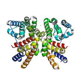
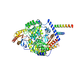
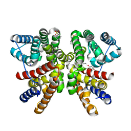
2BYB
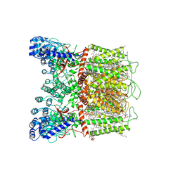
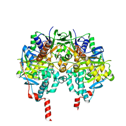 | | Human Monoamine Oxidase B in complex with Deprenyl | | 分子名称: | AMINE OXIDASE [FLAVIN-CONTAINING] B, DEPRENYL, FLAVIN-ADENINE DINUCLEOTIDE | | 著者 | Binda, C, De Colibus, L, Edmondson, D.E, Mattevi, A. | | 登録日 | 2005-07-29 | | 公開日 | 2005-08-09 | | 最終更新日 | 2023-12-13 | | 実験手法 | X-RAY DIFFRACTION (2.2 Å) | | 主引用文献 | Three-Dimensional Structure of Human Monoamine Oxidase a (Mao A): Relation to the Structures of Rat Mao a and Human Mao B
Proc.Natl.Acad.Sci.USA, 102, 2005
|
|
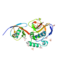
6FGE
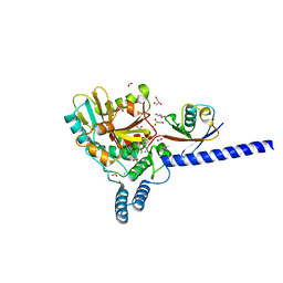 | | Crystal structure of human ZUFSP/ZUP1 in complex with ubiquitin | | 分子名称: | ACETATE ION, AMMONIUM ION, DI(HYDROXYETHYL)ETHER, ... | | 著者 | Kwasna, D, Abdul Rehman, S.A, Kulathu, Y. | | 登録日 | 2018-01-10 | | 公開日 | 2018-04-04 | | 最終更新日 | 2018-04-18 | | 実験手法 | X-RAY DIFFRACTION (1.74 Å) | | 主引用文献 | Discovery and Characterization of ZUFSP/ZUP1, a Distinct Deubiquitinase Class Important for Genome Stability.
Mol. Cell, 70, 2018
|
|
2BIW
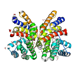
 | | Crystal structure of apocarotenoid cleavage oxygenase from Synechocystis, native enzyme | | 分子名称: | (3R)-3-HYDROXY-8'-APOCAROTENOL, APOCAROTENOID-CLEAVING OXYGENASE, FE (III) ION | | 著者 | Kloer, D.P, Ruch, S, Al-Babili, S, Beyer, P, Schulz, G.E. | | 登録日 | 2005-01-26 | | 公開日 | 2005-04-14 | | 最終更新日 | 2024-05-01 | | 実験手法 | X-RAY DIFFRACTION (2.39 Å) | | 主引用文献 | The Structure of a Retinal-Forming Carotenoid Oxygenase
Science, 308, 2005
|
|
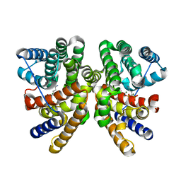
2CBQ
 | | Crystal structure of the neocarzinostatin 1Tes15 mutant bound to testosterone hemisuccinate. | | 分子名称: | NEOCARZINOSTATIN, SULFATE ION, TESTOSTERONE HEMISUCCINATE | | 著者 | Drevelle, A, Graille, M, Heyd, B, Sorel, I, Ulryck, N, Pecorari, F, Desmadril, M, Van Tilbeurgh, H, Minard, P. | | 登録日 | 2006-01-06 | | 公開日 | 2006-03-22 | | 最終更新日 | 2023-12-13 | | 実験手法 | X-RAY DIFFRACTION (2.6 Å) | | 主引用文献 | Structures of in Vitro Evolved Binding Sites on Neocarzinostatin Scaffold Reveal Unanticipated Evolutionary Pathways.
J.Mol.Biol., 358, 2006
|
|
6GGL
 | | Crystal structure of CotB2 variant F107A | | 分子名称: | CHLORIDE ION, Cyclooctat-9-en-7-ol synthase, MAGNESIUM ION | | 著者 | Driller, R, Janke, S, Fuchs, M, Warner, E, Mhashal, A.R, Major, D.T, Christmann, M, Brueck, T, Loll, B. | | 登録日 | 2018-05-03 | | 公開日 | 2018-10-10 | | 最終更新日 | 2024-01-17 | | 実験手法 | X-RAY DIFFRACTION (1.9 Å) | | 主引用文献 | Towards a comprehensive understanding of the structural dynamics of a bacterial diterpene synthase during catalysis.
Nat Commun, 9, 2018
|
|
6FFA
 | | FMDV Leader protease bound to substrate ISG15 | | 分子名称: | GLYCEROL, Lbpro, SULFATE ION, ... | | 著者 | Swatek, K.N, Pruneda, J.N, Komander, D. | | 登録日 | 2018-01-05 | | 公開日 | 2018-02-21 | | 最終更新日 | 2024-01-17 | | 実験手法 | X-RAY DIFFRACTION (1.5 Å) | | 主引用文献 | Irreversible inactivation of ISG15 by a viral leader protease enables alternative infection detection strategies.
Proc. Natl. Acad. Sci. U.S.A., 115, 2018
|
|
6GGI
 | | Crystal structure of CotB2 in complex with 2-fluoro-3,7,18-dolabellatriene | | 分子名称: | (4S)-2-METHYL-2,4-PENTANEDIOL, 2-fluoro-3,7,18-dolabellatriene, ACETONITRILE, ... | | 著者 | Driller, R, Janke, S, Fuchs, M, Warner, E, Mhashal, A.R, Major, D.T, Christmann, M, Brueck, T, Loll, B. | | 登録日 | 2018-05-03 | | 公開日 | 2018-10-10 | | 最終更新日 | 2024-01-17 | | 実験手法 | X-RAY DIFFRACTION (1.803 Å) | | 主引用文献 | Towards a comprehensive understanding of the structural dynamics of a bacterial diterpene synthase during catalysis.
Nat Commun, 9, 2018
|
|
6GGK
 | | Crystal structure of CotB2 C-terminal truncation | | 分子名称: | CHLORIDE ION, Cyclooctat-9-en-7-ol synthase, MAGNESIUM ION | | 著者 | Driller, R, Janke, S, Fuchs, M, Warner, E, Mhashal, A.R, Major, D.T, Christmann, M, Brueck, T, Loll, B. | | 登録日 | 2018-05-03 | | 公開日 | 2018-10-10 | | 最終更新日 | 2024-01-17 | | 実験手法 | X-RAY DIFFRACTION (2.15 Å) | | 主引用文献 | Towards a comprehensive understanding of the structural dynamics of a bacterial diterpene synthase during catalysis.
Nat Commun, 9, 2018
|
|
6GGJ
 | | Crystal structure of CotB2 in complex with alendronate | | 分子名称: | 4-AMINO-1-HYDROXYBUTANE-1,1-DIYLDIPHOSPHONATE, Cyclooctat-9-en-7-ol synthase, MAGNESIUM ION | | 著者 | Driller, R, Janke, S, Fuchs, M, Warner, E, Mhashal, A.R, Major, D.T, Christmann, M, Brueck, T, Loll, B. | | 登録日 | 2018-05-03 | | 公開日 | 2018-10-10 | | 最終更新日 | 2024-01-17 | | 実験手法 | X-RAY DIFFRACTION (2.1 Å) | | 主引用文献 | Towards a comprehensive understanding of the structural dynamics of a bacterial diterpene synthase during catalysis.
Nat Commun, 9, 2018
|
|
7K0K
 | | Human serine palmitoyltransferase complex SPTLC1/SPLTC2/ssSPTa, 3KS-bound | | 分子名称: | 3-Dehydrosphinganine, PYRIDOXAL-5'-PHOSPHATE, Serine palmitoyltransferase 1, ... | | 著者 | Wang, Y, Niu, Y, Zhang, Z, Zhao, H, Myasnikov, A, Kalathur, R, Lee, C.H. | | 登録日 | 2020-09-04 | | 公開日 | 2021-02-24 | | 最終更新日 | 2021-03-24 | | 実験手法 | ELECTRON MICROSCOPY (2.6 Å) | | 主引用文献 | Structural insights into the regulation of human serine palmitoyltransferase complexes.
Nat.Struct.Mol.Biol., 28, 2021
|
|
8PD0
 | | cryo-EM structure of Doa10 in MSP1E3D1 | | 分子名称: | 1,2-DIOLEOYL-SN-GLYCERO-3-PHOSPHOCHOLINE, 1,2-DIPALMITOYL-SN-GLYCERO-3-PHOSPHATE, ERAD-associated E3 ubiquitin-protein ligase DOA10 | | 著者 | Botsch, J.J, Braeuning, B, Schulman, B.A. | | 登録日 | 2023-06-11 | | 公開日 | 2024-01-17 | | 実験手法 | ELECTRON MICROSCOPY (3.58 Å) | | 主引用文献 | Doa10/MARCH6 architecture interconnects E3 ligase activity with lipid-binding transmembrane channel to regulate SQLE.
Nat Commun, 15, 2024
|
|
8PDA
 | | cryo-EM structure of Doa10 with RING domain in MSP1E3D1 | | 分子名称: | 1,2-DIOLEOYL-SN-GLYCERO-3-PHOSPHOCHOLINE, 1,2-DIPALMITOYL-SN-GLYCERO-3-PHOSPHATE, ERAD-associated E3 ubiquitin-protein ligase DOA10 | | 著者 | Botsch, J.J, Braeuning, B, Schulman, B.A. | | 登録日 | 2023-06-12 | | 公開日 | 2024-01-17 | | 実験手法 | ELECTRON MICROSCOPY (3.58 Å) | | 主引用文献 | Doa10/MARCH6 architecture interconnects E3 ligase activity with lipid-binding transmembrane channel to regulate SQLE.
Nat Commun, 15, 2024
|
|
7JJU
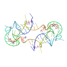 | | Crystal structure of en exoribonuclease-resistant RNA (xrRNA) from Potato leafroll virus (PLRV) | | 分子名称: | CACODYLATE ION, Guanidinium, IRIDIUM HEXAMMINE ION, ... | | 著者 | Steckelberg, A.-L, Vicens, Q, Auffinger, P, Costantino, D.C, Nix, J.C, Kieft, J.S. | | 登録日 | 2020-07-27 | | 公開日 | 2020-09-09 | | 最終更新日 | 2024-03-06 | | 実験手法 | X-RAY DIFFRACTION (2.604 Å) | | 主引用文献 | The crystal structure of a Polerovirus exoribonuclease-resistant RNA shows how diverse sequences are integrated into a conserved fold.
Rna, 26, 2020
|
|
8QWR
 | | Crystal structure of CotB2 variant V80L | | 分子名称: | (4S)-2-METHYL-2,4-PENTANEDIOL, 3-CYCLOHEXYL-1-PROPYLSULFONIC ACID, Cyclooctat-9-en-7-ol synthase, ... | | 著者 | Dimos, N, Himpich, S, Ringel, M, Driller, R, Major, D.T, Brueck, T, Loll, B. | | 登録日 | 2023-10-20 | | 公開日 | 2024-04-24 | | 実験手法 | X-RAY DIFFRACTION (1.7 Å) | | 主引用文献 | Crystal structure of CotB2 variant V80L
To Be Published
|
|
7K4B
 | | Cryo-EM structure of human TRPV6 in complex with (4- phenylcyclohexyl)piperazine inhibitor cis-22a | | 分子名称: | 1,2-DIOLEOYL-SN-GLYCERO-3-PHOSPHOCHOLINE, 1-[cis-4-(3-methylphenyl)cyclohexyl]-4-(pyridin-3-yl)piperazine, CALCIUM ION, ... | | 著者 | Neuberger, A, Nadezhdin, K.D, Singh, A.K, Sobolevsky, A.I. | | 登録日 | 2020-09-15 | | 公開日 | 2020-12-09 | | 最終更新日 | 2024-03-06 | | 実験手法 | ELECTRON MICROSCOPY (3.1 Å) | | 主引用文献 | Inactivation-mimicking block of the epithelial calcium channel TRPV6.
Sci Adv, 6, 2020
|
|
8QWS
 | | Crystal structure of CotB2 variant V80L in complex with alendronate | | 分子名称: | (4S)-2-METHYL-2,4-PENTANEDIOL, 1,2-ETHANEDIOL, 2-(2-METHOXYETHOXY)ETHANOL, ... | | 著者 | Dimos, N, Himpich, S, Ringel, M, Driller, R, Major, D.T, Brueck, T, Loll, B. | | 登録日 | 2023-10-20 | | 公開日 | 2024-04-24 | | 実験手法 | X-RAY DIFFRACTION (1.6 Å) | | 主引用文献 | Crystal structure of CotB2 variant V80L in complex with alendronate
To Be Published
|
|
8Q00
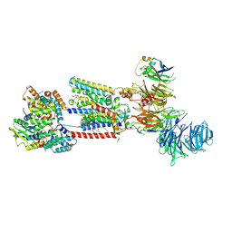
 | | TssM-Ub-PA complex - A USP-like DUB from B. pseudomallei (193-430) reacted with Ub-PA | | 分子名称: | 1,2-ETHANEDIOL, CITRATE ANION, Polyubiquitin-B, ... | | 著者 | Uthoff, M, Hermanns, T, Hofmann, K, Baumann, U. | | 登録日 | 2023-07-27 | | 公開日 | 2023-12-13 | | 最終更新日 | 2024-01-17 | | 実験手法 | X-RAY DIFFRACTION (1.62 Å) | | 主引用文献 | The structural basis for deubiquitination by the fingerless USP-type effector TssM.
Life Sci Alliance, 7, 2024
|
|
8S9S
 | | Structure of the human ER membrane protein complex (EMC) in GDN | | 分子名称: | 1,2-DIOLEOYL-SN-GLYCERO-3-PHOSPHOCHOLINE, 2-acetamido-2-deoxy-beta-D-glucopyranose, 2-acetamido-2-deoxy-beta-D-glucopyranose-(1-4)-2-acetamido-2-deoxy-beta-D-glucopyranose, ... | | 著者 | Tomaleri, G.P, Nguyen, V.N, Voorhees, R.M. | | 登録日 | 2023-03-30 | | 公開日 | 2023-05-17 | | 最終更新日 | 2023-06-14 | | 実験手法 | ELECTRON MICROSCOPY (3.6 Å) | | 主引用文献 | A selectivity filter in the ER membrane protein complex limits protein misinsertion at the ER.
J.Cell Biol., 222, 2023
|
|
8SLY
 | | Rat TRPV2 bound with 2 CBD ligands in nanodiscs | | 分子名称: | (2S)-3-(hexadecanoyloxy)-2-[(9Z)-octadec-9-enoyloxy]propyl 2-(trimethylammonio)ethyl phosphate, SODIUM ION, Transient receptor potential cation channel subfamily V member 2, ... | | 著者 | Tan, X, Swartz, K.J. | | 登録日 | 2023-04-24 | | 公開日 | 2023-05-31 | | 最終更新日 | 2024-06-19 | | 実験手法 | ELECTRON MICROSCOPY (3.32 Å) | | 主引用文献 | Cannabidiol sensitizes TRPV2 channels to activation by 2-APB.
Elife, 12, 2023
|
|
8SLX
 | |
7K4A
 | | Cryo-EM structure of human TRPV6 in the open state | | 分子名称: | 1,2-DIOLEOYL-SN-GLYCERO-3-PHOSPHOCHOLINE, CALCIUM ION, CHOLESTEROL HEMISUCCINATE, ... | | 著者 | Neuberger, A, Nadezhdin, K.D, Singh, A.K, Sobolevsky, A.I. | | 登録日 | 2020-09-15 | | 公開日 | 2020-12-09 | | 最終更新日 | 2024-03-06 | | 実験手法 | ELECTRON MICROSCOPY (3.26 Å) | | 主引用文献 | Inactivation-mimicking block of the epithelial calcium channel TRPV6.
Sci Adv, 6, 2020
|
|
8S9B
 | | Cryo-EM structure of Nav1.7 with LCM | | 分子名称: | 1,2-DIOLEOYL-SN-GLYCERO-3-PHOSPHOCHOLINE, 1-O-OCTADECYL-SN-GLYCERO-3-PHOSPHOCHOLINE, 2-acetamido-2-deoxy-beta-D-glucopyranose, ... | | 著者 | Fan, X, Huang, J, Yan, N. | | 登録日 | 2023-03-27 | | 公開日 | 2023-08-30 | | 実験手法 | ELECTRON MICROSCOPY (2.9 Å) | | 主引用文献 | Structural mapping of Na v 1.7 antagonists.
Nat Commun, 14, 2023
|
|
8S9C
 | | Cryo-EM structure of Nav1.7 with CBZ | | 分子名称: | 1,2-DIOLEOYL-SN-GLYCERO-3-PHOSPHOCHOLINE, 1-O-OCTADECYL-SN-GLYCERO-3-PHOSPHOCHOLINE, 2-acetamido-2-deoxy-beta-D-glucopyranose, ... | | 著者 | Fan, X, Huang, J, Yan, N. | | 登録日 | 2023-03-27 | | 公開日 | 2023-08-30 | | 実験手法 | ELECTRON MICROSCOPY (3.2 Å) | | 主引用文献 | Structural mapping of Na v 1.7 antagonists.
Nat Commun, 14, 2023
|
|


