8BUW
 
 | |
5VV2
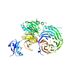
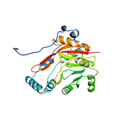
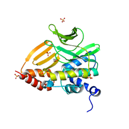
 | | Structure of human neuronal nitric oxide synthase heme domain in complex with 7-(((3-(5-Fluoropyridin-3-yl)propyl)amino)methyl)quinolin-2-amine | | 分子名称: | 5,6,7,8-TETRAHYDROBIOPTERIN, 7-({[3-(5-fluoropyridin-3-yl)propyl]amino}methyl)quinolin-2-amine, Nitric oxide synthase, ... | | 著者 | Huiying, L, Thomas, L.P. | | 登録日 | 2017-05-19 | | 公開日 | 2017-08-23 | | 最終更新日 | 2023-10-04 | | 実験手法 | X-RAY DIFFRACTION (2 Å) | | 主引用文献 | Hydrophilic, Potent, and Selective 7-Substituted 2-Aminoquinolines as Improved Human Neuronal Nitric Oxide Synthase Inhibitors.
J. Med. Chem., 60, 2017
|
|
5VWC
 | |
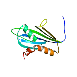
5VVF
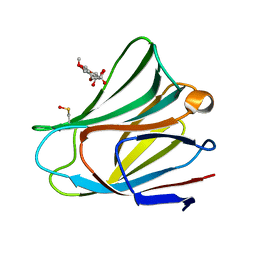
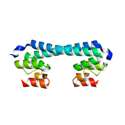
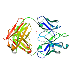 | | Crystal Structure of 354BG1 Fab | | 分子名称: | 1,2-ETHANEDIOL, 354BG1 Heavy Chain, 354BG1 Light Chain | | 著者 | Scharf, L, Gristick, H.B, Bjorkman, P.J. | | 登録日 | 2017-05-19 | | 公開日 | 2017-12-06 | | 最終更新日 | 2023-10-04 | | 実験手法 | X-RAY DIFFRACTION (2 Å) | | 主引用文献 | Asymmetric recognition of HIV-1 Envelope trimer by V1V2 loop-targeting antibodies.
Elife, 6, 2017
|
|
8BTX
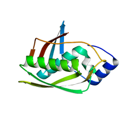 | |
7NBV
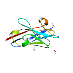
 | | Structure of 2A protein from Theilers murine encephalomyelitis virus (TMEV) | | 分子名称: | BROMIDE ION, Capsid protein VP0 | | 著者 | Hill, C.H, Cook, G.M, Napthine, S, Kibe, A, Brown, K, Caliskan, N, Firth, A.E, Graham, S.C, Brierley, I. | | 登録日 | 2021-01-28 | | 公開日 | 2021-12-08 | | 最終更新日 | 2024-06-19 | | 実験手法 | X-RAY DIFFRACTION (1.87 Å) | | 主引用文献 | Investigating molecular mechanisms of 2A-stimulated ribosomal pausing and frameshifting in Theilovirus.
Nucleic Acids Res., 49, 2021
|
|
7PO6
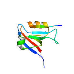
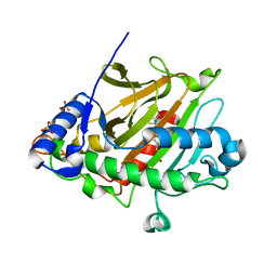
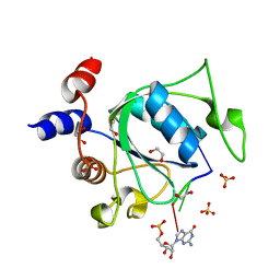 | | Xist (m6A)UCG tetraloop RNA bound to the YTH domain of YTHDC1 | | 分子名称: | 1,2-ETHANEDIOL, GUANOSINE-5'-TRIPHOSPHATE, Isoform 2 of YTH domain-containing protein 1, ... | | 著者 | Jones, A.N, Mourao, A, Sattler, M. | | 登録日 | 2021-09-08 | | 公開日 | 2022-03-16 | | 最終更新日 | 2024-01-31 | | 実験手法 | X-RAY DIFFRACTION (1.77 Å) | | 主引用文献 | Structural effects of m6A modification of the Xist A-repeat AUCG tetraloop and its recognition by YTHDC1.
Nucleic Acids Res., 50, 2022
|
|
5KMC
 | |
5EM2
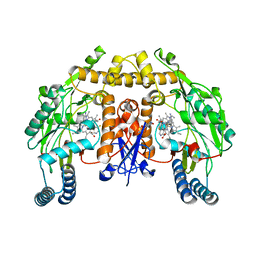
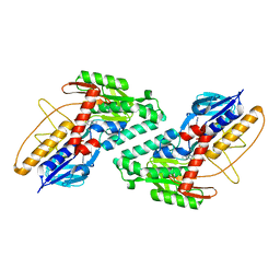
 | | Crystal structure of the Erb1-Ytm1 complex | | 分子名称: | 1,2-ETHANEDIOL, MAGNESIUM ION, Ribosome biogenesis protein ERB1, ... | | 著者 | Ahmed, Y.L, Sinning, I. | | 登録日 | 2015-11-05 | | 公開日 | 2015-12-23 | | 最終更新日 | 2024-05-08 | | 実験手法 | X-RAY DIFFRACTION (2.67 Å) | | 主引用文献 | Concerted removal of the Erb1-Ytm1 complex in ribosome biogenesis relies on an elaborate interface.
Nucleic Acids Res., 44, 2016
|
|
8BSY
 | |
5VWG
 | |
5KMQ
 | |
7PTZ
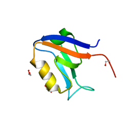
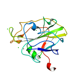 | | High resolution X-ray structure of E. coli expressed Lentinus similis LPMO. | | 分子名称: | Auxiliary activity 9, CHLORIDE ION, COPPER (II) ION | | 著者 | Banerjee, S, Muderspach, S.J, Tandrup, T, Ipsen, J.O, Hernandez-Rollan, C, Norholm, H.H.M, Johansen, K.S, Lo Leggio, L. | | 登録日 | 2021-09-27 | | 公開日 | 2022-03-16 | | 最終更新日 | 2024-01-31 | | 実験手法 | X-RAY DIFFRACTION (1.093 Å) | | 主引用文献 | Protonation State of an Important Histidine from High Resolution Structures of Lytic Polysaccharide Monooxygenases.
Biomolecules, 12, 2022
|
|
8BVI
 | |
8K48
 | |
8JZA
 | | Crystal structure of single domain antibody kt75 of human Thyroglobulin | | 分子名称: | ACETATE ION, POTASSIUM ION, single domain antibody kt75 of human Thyroglobulin | | 著者 | Mistry, H, Kumarasamy, J, Kumari, S, Kulkarni, S.P, Gupta, G.D. | | 登録日 | 2023-07-04 | | 公開日 | 2024-07-10 | | 実験手法 | X-RAY DIFFRACTION (1.35 Å) | | 主引用文献 | Crystal structure of single domain antibody kt75 of human Thyroglobulin
To Be Published
|
|
5ENF
 | | Crystal structure of the second bromodomain of Pleckstrin homology domain interacting protein (PHIP) in complex with fragment-4 N10142 (SGC - Diamond I04-1 fragment screening) | | 分子名称: | 1,2-ETHANEDIOL, 5-azanyl-2-(2-methylpropyl)-1,3-oxazole-4-carbonitrile, PH-interacting protein | | 著者 | Krojer, T, Talon, R, Collins, P, Bradley, A, Cox, O, Amin, J, Szykowska, A, Burgess-Brown, N, Spencer, J, Brennan, P, Bountra, C, Arrowsmith, C.H, Edwards, A, von Delft, F, Structural Genomics Consortium (SGC) | | 登録日 | 2015-11-09 | | 公開日 | 2016-04-27 | | 最終更新日 | 2024-01-10 | | 実験手法 | X-RAY DIFFRACTION (1.37 Å) | | 主引用文献 | A poised fragment library enables rapid synthetic expansion yielding the first reported inhibitors of PHIP(2), an atypical bromodomain.
Chem Sci, 7, 2016
|
|
8BSX
 | |
8K51
 | |
7NA9
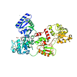
 | | Crystal structure of BoNT/B-LC-JSG-C1 | | 分子名称: | 1,2-ETHANEDIOL, Botulinum neurotoxin type B, JSG-C1, ... | | 著者 | Lam, K, Jin, R. | | 登録日 | 2021-06-20 | | 公開日 | 2021-12-22 | | 最終更新日 | 2023-10-18 | | 実験手法 | X-RAY DIFFRACTION (1.76 Å) | | 主引用文献 | Probing the structure and function of the protease domain of botulinum neurotoxins using single-domain antibodies.
Plos Pathog., 18, 2022
|
|
7Q1L
 | | Glycosilated Human Serum Apo-tranferrin | | 分子名称: | 1,2-ETHANEDIOL, 2-acetamido-2-deoxy-beta-D-glucopyranose-(1-4)-2-acetamido-2-deoxy-beta-D-glucopyranose, GLYCEROL, ... | | 著者 | Gavira, J.A, Moreno, A, Campos-Escamilla, C, Gonzalez-Ramirez, L.A, Siliqi, D. | | 登録日 | 2021-10-20 | | 公開日 | 2022-03-16 | | 最終更新日 | 2024-01-31 | | 実験手法 | X-RAY DIFFRACTION (3 Å) | | 主引用文献 | X-ray Characterization of Conformational Changes of Human Apo- and Holo-Transferrin.
Int J Mol Sci, 22, 2021
|
|
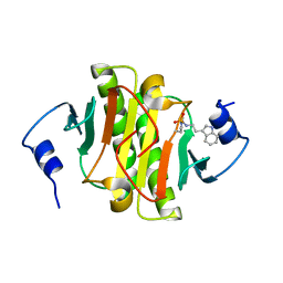
7NIT
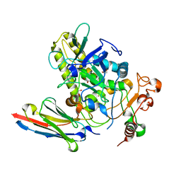
 | | X-ray structure of a multidomain BbgIII from Bifidobacterium bifidum | | 分子名称: | Beta-galactosidase, CALCIUM ION, GLYCEROL, ... | | 著者 | Moroz, O.V, Blagova, E, Lebedev, A.A, Sanchez Rodriguez, F, Rigden, D.J, Tams, J.W, Wilting, R, Vester, J.K, Longhin, E, Krogh, K.B.R, Pache, R.A, Davies, G.J, Wilson, K.S. | | 登録日 | 2021-02-14 | | 公開日 | 2021-12-22 | | 最終更新日 | 2024-01-31 | | 実験手法 | X-RAY DIFFRACTION (2.89 Å) | | 主引用文献 | Multitasking in the gut: the X-ray structure of the multidomain BbgIII from Bifidobacterium bifidum offers possible explanations for its alternative functions.
Acta Crystallogr D Struct Biol, 77, 2021
|
|
5EH5
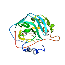 | | human carbonic anhydrase II in complex with ligand | | 分子名称: | 1.7.6 4-methylpyrimidine-2-sulfonamide, Carbonic anhydrase 2, FORMIC ACID, ... | | 著者 | Ren, B. | | 登録日 | 2015-10-28 | | 公開日 | 2016-03-09 | | 最終更新日 | 2024-03-06 | | 実験手法 | X-RAY DIFFRACTION (1.207 Å) | | 主引用文献 | Native State Mass Spectrometry, Surface Plasmon Resonance, and X-ray Crystallography Correlate Strongly as a Fragment Screening Combination.
J.Med.Chem., 59, 2016
|
|
7VHC
 | | Crystal structure of the STX2a complexed with AR4A peptide | | 分子名称: | 3-PYRIDINIUM-1-YLPROPANE-1-SULFONATE, Shiga toxin 2 B subunit, inhibitor peptide, ... | | 著者 | Senda, M, Takahashi, M, Nishikawa, K, Senda, T. | | 登録日 | 2021-09-22 | | 公開日 | 2022-07-20 | | 最終更新日 | 2023-11-29 | | 実験手法 | X-RAY DIFFRACTION (1.8 Å) | | 主引用文献 | A unique peptide-based pharmacophore identifies an inhibitory compound against the A-subunit of Shiga toxin.
Sci Rep, 12, 2022
|
|
7NHF
 | | Crystal structure of Arabidopsis thaliana Pdx1K166R | | 分子名称: | PHOSPHATE ION, Pyridoxal 5'-phosphate synthase subunit PDX1.3 | | 著者 | Rodrigues, M.J, Zhang, Y, Bolton, R, Evans, G, Giri, N, Royant, A, Begley, T, Ealick, S.E, Tews, I. | | 登録日 | 2021-02-10 | | 公開日 | 2021-12-22 | | 最終更新日 | 2024-01-31 | | 実験手法 | X-RAY DIFFRACTION (2.35 Å) | | 主引用文献 | Trapping and structural characterisation of a covalent intermediate in vitamin B 6 biosynthesis catalysed by the Pdx1 PLP synthase.
Rsc Chem Biol, 3, 2022
|
|


