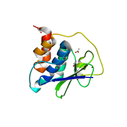
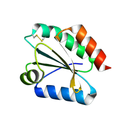
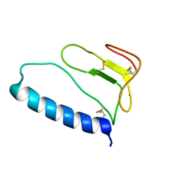
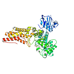
1ZT3
 
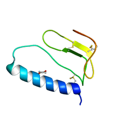 | | C-terminal domain of Insulin-like Growth Factor Binding Protein-1 isolated from human amniotic fluid | | 分子名称: | 1,4-DIETHYLENE DIOXIDE, Insulin-like growth factor binding protein 1 | | 著者 | Sala, A, Capaldi, S, Campagnoli, M, Faggion, B, Labo, S, Perduca, M, Romano, A, Carrizo, M.E, Valli, M, Visai, L, Minchiotti, L, Galliano, M, Monaco, H.L. | | 登録日 | 2005-05-26 | | 公開日 | 2005-06-28 | | 最終更新日 | 2017-10-11 | | 実験手法 | X-RAY DIFFRACTION (1.8 Å) | | 主引用文献 | Structure and Properties of the C-terminal Domain of Insulin-like Growth Factor-binding Protein-1 Isolated from Human Amniotic Fluid
J.Biol.Chem., 280, 2005
|
|
4YZY
 | |
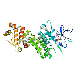
4TZ7
 | | Crystal structure of type I phosphatidylinositol 4-phosphate 5-kinase alpha from Zebrafish | | 分子名称: | Phosphatidylinositol-4-phosphate 5-kinase, type I, alpha | | 著者 | Hu, J, Qin, Y, Wang, J, Li, L, Wu, D, Ha, Y. | | 登録日 | 2014-07-09 | | 公開日 | 2015-09-02 | | 最終更新日 | 2023-12-27 | | 実験手法 | X-RAY DIFFRACTION (3.31 Å) | | 主引用文献 | Resolution of structure of PIP5K1A reveals molecular mechanism for its regulation by dimerization and dishevelled.
Nat Commun, 6, 2015
|
|
1YGO
 | |
1CHQ
 | |
2PYO
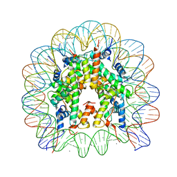 | | Drosophila nucleosome core | | 分子名称: | CHLORIDE ION, DNA (147-MER), Histone H2A, ... | | 著者 | Clapier, C.R, Petosa, C, Mueller, C.W. | | 登録日 | 2007-05-16 | | 公開日 | 2007-11-06 | | 最終更新日 | 2024-02-21 | | 実験手法 | X-RAY DIFFRACTION (2.43 Å) | | 主引用文献 | Structure of the Drosophila nucleosome core particle highlights evolutionary constraints on the H2A-H2B histone dimer.
Proteins, 71, 2007
|
|
1Y9I
 | | Crystal structure of low temperature requirement C protein from Listeria monocytogenes | | 分子名称: | CALCIUM ION, GLYCEROL, MAGNESIUM ION, ... | | 著者 | Kumaran, D, Swaminathan, S, Burley, S.K, New York SGX Research Center for Structural Genomics (NYSGXRC) | | 登録日 | 2004-12-15 | | 公開日 | 2004-12-28 | | 最終更新日 | 2021-02-03 | | 実験手法 | X-RAY DIFFRACTION (1.8 Å) | | 主引用文献 | Crystal structure of phosphatidylglycerophosphatase (PGPase), a putative membrane-bound lipid phosphatase, reveals a novel binuclear metal binding site and two "proton wires".
Proteins, 64, 2006
|
|
1CHP
 | |
8EHO
 | | PRRSV-1 PLP2 domain bound to ubiquitin | | 分子名称: | 3-AMINOPROPANE, GLYCEROL, NITRATE ION, ... | | 著者 | Bailey-Elkin, B.A, Mark, B.L. | | 登録日 | 2022-09-14 | | 公開日 | 2023-12-06 | | 最終更新日 | 2024-01-03 | | 実験手法 | X-RAY DIFFRACTION (2.85 Å) | | 主引用文献 | Demonstrating the importance of porcine reproductive and respiratory syndrome virus papain-like protease 2 deubiquitinating activity in viral replication by structure-guided mutagenesis.
Plos Pathog., 19, 2023
|
|
8EHN
 | | PRRSV-1 PLP2 domain | | 分子名称: | ACETATE ION, Papain-like protease 2, ZINC ION | | 著者 | Bailey-Elkin, B.A, Mark, B.L. | | 登録日 | 2022-09-14 | | 公開日 | 2023-12-06 | | 最終更新日 | 2024-01-03 | | 実験手法 | X-RAY DIFFRACTION (2.3 Å) | | 主引用文献 | Demonstrating the importance of porcine reproductive and respiratory syndrome virus papain-like protease 2 deubiquitinating activity in viral replication by structure-guided mutagenesis.
Plos Pathog., 19, 2023
|
|
4YZD
 | |
4RQK
 | |
1TCB
 | |
2VM2
 | | Crystal structure of barley thioredoxin h isoform 1 crystallized using PEG as precipitant | | 分子名称: | THIOREDOXIN H ISOFORM 1. | | 著者 | Maeda, K, Hagglund, P, Finnie, C, Svensson, B, Henriksen, A. | | 登録日 | 2008-01-21 | | 公開日 | 2008-04-29 | | 最終更新日 | 2023-12-13 | | 実験手法 | X-RAY DIFFRACTION (1.8 Å) | | 主引用文献 | Crystal Structures of Barley Thioredoxin H Isoforms Hvtrxh1 and Hvtrxh2 Reveal Features Involved in Protein Recognition and Possibly in Discriminating the Isoform Specificity.
Protein Sci., 17, 2008
|
|
1YI5
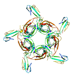 | | Crystal structure of the a-cobratoxin-AChBP complex | | 分子名称: | Acetylcholine-binding protein, Long neurotoxin 1 | | 著者 | Bourne, Y, Talley, T.T, Hansen, S.B, Taylor, P, Marchot, P. | | 登録日 | 2005-01-11 | | 公開日 | 2005-05-17 | | 最終更新日 | 2023-10-25 | | 実験手法 | X-RAY DIFFRACTION (4.2 Å) | | 主引用文献 | Crystal structure of a Cbtx-AChBP complex reveals essential interactions between snake alpha-neurotoxins and nicotinic receptors
Embo J., 24, 2005
|
|
4XYN
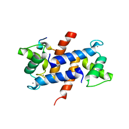 | | X-ray structure of Ca(2+)-S100B with human RAGE-derived W61 peptide | | 分子名称: | CALCIUM ION, Protein S100-B, Receptor for advanced glycation endproducts-derived peptide (W61) | | 著者 | Jensen, J.L, Indurthi, V.S.K, Neau, D, Vetter, S.W, Colbert, C.L. | | 登録日 | 2015-02-02 | | 公開日 | 2015-05-13 | | 最終更新日 | 2024-02-28 | | 実験手法 | X-RAY DIFFRACTION (2.55 Å) | | 主引用文献 | Structural insights into the binding of the human receptor for advanced glycation end products (RAGE) by S100B, as revealed by an S100B-RAGE-derived peptide complex.
Acta Crystallogr.,Sect.D, 71, 2015
|
|
1ZR0
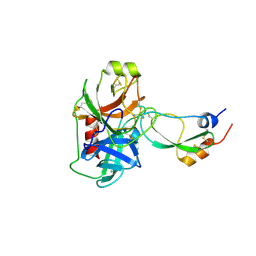 | | Crystal Structure of Kunitz Domain 1 of Tissue Factor Pathway Inhibitor-2 with Bovine Trypsin | | 分子名称: | CALCIUM ION, Cationic trypsin, Tissue factor pathway inhibitor 2 | | 著者 | Schmidt, A.E, Chand, H.S, Cascio, D, Kisiel, W, Bajaj, S.P. | | 登録日 | 2005-05-18 | | 公開日 | 2005-06-07 | | 最終更新日 | 2023-08-23 | | 実験手法 | X-RAY DIFFRACTION (1.802 Å) | | 主引用文献 | Crystal structure of Kunitz domain 1 (KD1) of tissue factor pathway inhibitor-2 in complex with trypsin. Implications for KD1 specificity of inhibition
J.Biol.Chem., 280, 2005
|
|
1ZT5
 | | C-terminal domain of Insulin-like Growth Factor Binding Protein-1 isolated from human amniotic fluid complexed with Iron(II) | | 分子名称: | 1,4-DIETHYLENE DIOXIDE, FE (II) ION, Insulin-like growth factor binding protein 1 | | 著者 | Sala, A, Capaldi, S, Campagnoli, M, Faggion, B, Labo, S, Perduca, M, Romano, A, Carrizo, M.E, Valli, M, Visai, L, Minchiotti, L, Galliano, M, Monaco, H.L. | | 登録日 | 2005-05-26 | | 公開日 | 2005-06-28 | | 最終更新日 | 2011-07-13 | | 実験手法 | X-RAY DIFFRACTION (1.818 Å) | | 主引用文献 | Structure and Properties of the C-terminal Domain of Insulin-like Growth Factor-binding Protein-1 Isolated from Human Amniotic Fluid
J.Biol.Chem., 280, 2005
|
|
1ZU1
 | | Solution Structure of the N-terminal Zinc Fingers of the Xenopus laevis double stranded RNA binding protein ZFa | | 分子名称: | RNA binding protein ZFa, ZINC ION | | 著者 | Moller, H.M, Martinez-Yamout, M.A, Dyson, H.J, Wright, P.E. | | 登録日 | 2005-05-29 | | 公開日 | 2005-09-20 | | 最終更新日 | 2024-05-22 | | 実験手法 | SOLUTION NMR | | 主引用文献 | Solution structure of the N-terminal zinc fingers of the Xenopus laevis double-stranded RNA-binding protein ZFa
J.Mol.Biol., 351, 2005
|
|
1BKJ
 | | NADPH:FMN OXIDOREDUCTASE FROM VIBRIO HARVEYI | | 分子名称: | FLAVIN MONONUCLEOTIDE, NADPH-FLAVIN OXIDOREDUCTASE, PHOSPHATE ION | | 著者 | Tanner, J.J, Lei, B, TU, S.-C, Krause, K.L. | | 登録日 | 1998-07-08 | | 公開日 | 1999-01-13 | | 最終更新日 | 2024-02-07 | | 実験手法 | X-RAY DIFFRACTION (1.8 Å) | | 主引用文献 | Flavin reductase P: structure of a dimeric enzyme that reduces flavin.
Biochemistry, 35, 1996
|
|
1V5F
 | | Crystal Structure of Pyruvate oxidase complexed with FAD and TPP, from Aerococcus viridans | | 分子名称: | FLAVIN-ADENINE DINUCLEOTIDE, MAGNESIUM ION, Pyruvate oxidase, ... | | 著者 | Hossain, M.T, Suzuki, K, Yamamoto, T, Imamura, S, Sekiguchi, T, Takenaka, A. | | 登録日 | 2003-11-22 | | 公開日 | 2005-06-28 | | 最終更新日 | 2023-10-25 | | 実験手法 | X-RAY DIFFRACTION (1.8 Å) | | 主引用文献 | The structures of pyruvate oxidase from Aerococcus viridans with cofactors and with a reaction intermediate reveal the flexibility of the active-site tunnel for catalysis.
Acta Crystallogr.,Sect.F, 63, 2007
|
|
2W4X
 | | BtGH84 in complex with STZ | | 分子名称: | CALCIUM ION, GLYCEROL, O-GLCNACASE BT_4395, ... | | 著者 | He, Y, Bubb, A, Martinez-Fleites, C, Davies, G.J. | | 登録日 | 2008-12-02 | | 公開日 | 2009-02-24 | | 最終更新日 | 2023-12-13 | | 実験手法 | X-RAY DIFFRACTION (2.42 Å) | | 主引用文献 | Structural Insight Into the Mechanism of Streptozotocin Inhibition of O-Glcnacase.
Carbohydr.Res., 344, 2009
|
|
2PO3
 | | Crystal Structure Analysis of DesI in the presence of its TDP-sugar product | | 分子名称: | (2R,3R,4S,5S,6R)-3,4-DIHYDROXY-5-[({3-HYDROXY-2-METHYL-5-[(PHOSPHONOOXY)METHYL]PYRIDIN-4-YL}METHYL)IMINO]-6-METHYLTETRAHYDRO-2H-PYRAN-2-YL [(2R,3S,5R)-3-HYDROXY-5-(5-METHYL-2,4-DIOXO-3,4-DIHYDROPYRIMIDIN-1(2H)-YL)TETRAHYDROFURAN-2-YL]METHYL DIHYDROGEN DIPHOSPHATE, 4-dehydrase | | 著者 | Burgie, E.S, Holden, H.M. | | 登録日 | 2007-04-25 | | 公開日 | 2007-08-14 | | 最終更新日 | 2023-08-30 | | 実験手法 | X-RAY DIFFRACTION (2.1 Å) | | 主引用文献 | Molecular Architecture of DesI: A Key Enzyme in the Biosynthesis of Desosamine
Biochemistry, 46, 2007
|
|
8QU9
 | | Structure of the NCOA4 (Nuclear Receptor Coactivator 4)-FTH1 (H-Ferritin) complex | | 分子名称: | FE (III) ION, Ferritin heavy chain, NCOA4 (Nuclear Receptor Coactivator 4) | | 著者 | Hoelzgen, F, Klukin, E, Zalk, R, Shahar, A, Cohen-Schwartz, S, Frank, G.A. | | 登録日 | 2023-10-15 | | 公開日 | 2024-05-15 | | 実験手法 | ELECTRON MICROSCOPY (2.88 Å) | | 主引用文献 | Structural basis for the intracellular regulation of ferritin degradation.
Nat Commun, 15, 2024
|
|
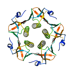
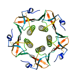
8FFD
 | | Crystal structure of manganeese bound Dps protein (PA0962) from Pseudomonas aeruginosa (cubic form) | | 分子名称: | L(+)-TARTARIC ACID, MANGANESE (II) ION, Probable dna-binding stress protein | | 著者 | Lovell, S, Seibold, S, Battaile, K.P, Rivera, M. | | 登録日 | 2022-12-08 | | 公開日 | 2023-03-08 | | 最終更新日 | 2024-05-22 | | 実験手法 | X-RAY DIFFRACTION (2.2 Å) | | 主引用文献 | Pseudomonas aeruginosa Dps (PA0962) Functions in H 2 O 2 Mediated Oxidative Stress Defense and Exhibits In Vitro DNA Cleaving Activity.
Int J Mol Sci, 24, 2023
|
|


