Loading
PDBj
メニューPDBj@FacebookPDBj@TwitterPDBj@YouTubewwPDB FoundationwwPDB
RCSB PDBPDBeBMRBAdv. SearchSearch help

2H1E
DownloadVisualize
BU of 2h1e by Molmil
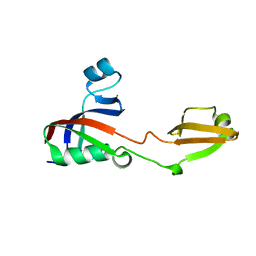
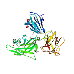
Tandem chromodomains of budding yeast CHD1
分子名称: Chromo domain protein 1
著者Flanagan IV, J.F, Khorasanizadeh, S.
登録日2006-05-16
公開日2007-03-27
最終更新日2023-08-30
実験手法X-RAY DIFFRACTION (2.2 Å)
主引用文献Molecular Implications of Evolutionary Differences in CHD Double Chromodomains.
J.Mol.Biol., 369, 2007
3PEY
DownloadVisualize
BU of 3pey by Molmil
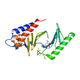
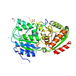
S. cerevisiae Dbp5 bound to RNA and ADP BeF3
分子名称: ADENOSINE-5'-DIPHOSPHATE, ATP-dependent RNA helicase DBP5, BERYLLIUM TRIFLUORIDE ION, ...
著者Montpetit, B, Thomsen, N.D, Helmke, K.J, Seeliger, M.A, Berger, J.M, Weis, K.
登録日2010-10-27
公開日2011-03-23
最終更新日2024-02-21
実験手法X-RAY DIFFRACTION (1.401 Å)
主引用文献A conserved mechanism of DEAD-box ATPase activation by nucleoporins and InsP(6) in mRNA export.
Nature, 472, 2011
2ESO
DownloadVisualize
BU of 2eso by Molmil
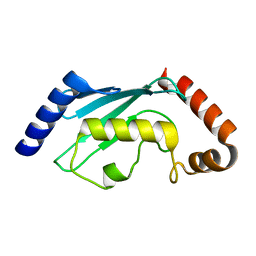
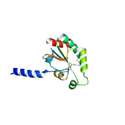
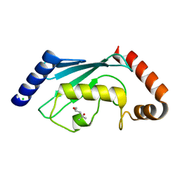
Human Ubiquitin-Conjugating Enzyme (E2) UbcH5b mutant Ile37Ala
分子名称: Ubiquitin-conjugating enzyme E2 D2
著者Ozkan, E, Yu, H, Deisenhofer, J.
登録日2005-10-26
公開日2005-12-06
最終更新日2023-08-23
実験手法X-RAY DIFFRACTION (1.5 Å)
主引用文献Mechanistic insight into the allosteric activation of a ubiquitin-conjugating enzyme by RING-type ubiquitin ligases
Proc.Natl.Acad.Sci.Usa, 102, 2005
2R2H
DownloadVisualize
BU of 2r2h by Molmil
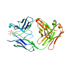
Structure of S25-2 in Complex with Ko
分子名称: Fab, antibody fragment (IgG1k), heavy chain, ...
著者Brooks, C.L, Evans, S.V.
登録日2007-08-25
公開日2008-12-30
最終更新日2020-07-29
実験手法X-RAY DIFFRACTION (2 Å)
主引用文献Exploration of specificity in germline monoclonal antibody recognition of a range of natural and synthetic epitopes.
J.Mol.Biol., 377, 2008
3PPH
DownloadVisualize
BU of 3pph by Molmil
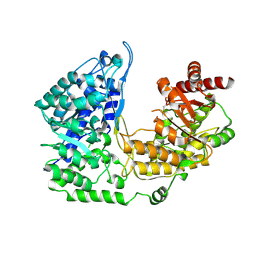
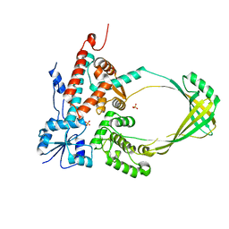
Crystal structure of the Candida albicans methionine synthase by surface entropy reduction, threonine variant
分子名称: 5-methyltetrahydropteroyltriglutamate--homocysteine methyltransferase
著者Ubhi, D, Kavanagh, K, Monzingo, A.F, Robertus, J.D.
登録日2010-11-24
公開日2011-10-12
最終更新日2023-09-06
実験手法X-RAY DIFFRACTION (2.8 Å)
主引用文献Structure of Candida albicans methionine synthase determined by employing surface residue mutagenesis.
Arch.Biochem.Biophys., 513, 2011
3PR9
DownloadVisualize
BU of 3pr9 by Molmil
Structural analysis of protein folding by the Methanococcus jannaschii chaperone FKBP26
分子名称: FKBP-type peptidyl-prolyl cis-trans isomerase
著者Martinez-Hackert, E, Hendrickson, W.A.
登録日2010-11-29
公開日2011-01-19
最終更新日2024-02-21
実験手法X-RAY DIFFRACTION (1.95 Å)
主引用文献Structural Analysis of Protein Folding by the Long-Chain Archaeal Chaperone FKBP26.
J.Mol.Biol., 407, 2011
4P6A
DownloadVisualize
BU of 4p6a by Molmil
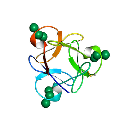
Crystal structure of a potent anti-HIV lectin actinohivin in complex with alpha-1,2-mannotriose
分子名称: Actinohivin, alpha-D-mannopyranose-(1-2)-alpha-D-mannopyranose-(1-2)-alpha-D-mannopyranose
著者Zhang, F, Hoque, M.M, Suzuki, K, Tsunoda, M, Naomi, O, Tanaka, H, Takenaka, A.
登録日2014-03-23
公開日2015-03-04
最終更新日2023-12-27
実験手法X-RAY DIFFRACTION (1.398 Å)
主引用文献The characteristic structure of anti-HIV actinohivin in complex with three HMTG D1 chains of HIV-gp120.
Chembiochem, 15, 2014
3HA9
DownloadVisualize
BU of 3ha9 by Molmil
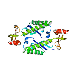
The 1.7A Crystal Structure of a Thioredoxin-like Protein from Aeropyrum pernix
分子名称: uncharacterized Thioredoxin-like protein
著者Stein, A.J, Cuff, M.E, Sather, A, Hendricks, R, Freeman, L, Joachimiak, A, Midwest Center for Structural Genomics (MCSG)
登録日2009-05-01
公開日2009-05-19
最終更新日2017-11-01
実験手法X-RAY DIFFRACTION (1.7 Å)
主引用文献The 1.7A Crystal Structure of a Thioredoxin-like Protein from Aeropyrum pernix
To be Published
4I1A
DownloadVisualize
BU of 4i1a by Molmil
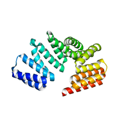
Crystal Structure of the Apo Form of RapI
分子名称: CHLORIDE ION, Response regulator aspartate phosphatase I
著者Parashar, V, Jeffrey, P.D, Neiditch, M.B.
登録日2012-11-20
公開日2013-04-03
最終更新日2024-02-28
実験手法X-RAY DIFFRACTION (2.443 Å)
主引用文献Conformational change-induced repeat domain expansion regulates rap phosphatase quorum-sensing signal receptors.
Plos Biol., 11, 2013
2QDC
DownloadVisualize
BU of 2qdc by Molmil
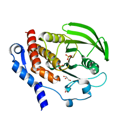
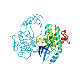
Crystal structure of the HePTP catalytic domain D236A mutant
分子名称: GLYCEROL, PHOSPHATE ION, Tyrosine-protein phosphatase non-receptor type 7
著者Critton, D.A, Tortajada, A, Page, R.
登録日2007-06-20
公開日2008-06-24
最終更新日2023-09-20
実験手法X-RAY DIFFRACTION (2 Å)
主引用文献Structural basis of substrate recognition by hematopoietic tyrosine phosphatase.
Biochemistry, 47, 2008
4I1W
DownloadVisualize
BU of 4i1w by Molmil
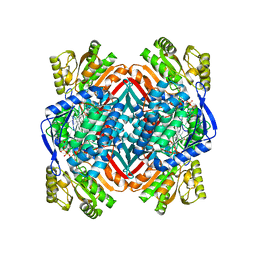
2.00 Angstroms X-ray crystal structure of NAD- bound 2-aminomuconate 6-semialdehyde dehydrogenase from Pseudomonas fluorescens
分子名称: 2-aminomuconate 6-semialdehyde dehydrogenase, GLYCEROL, NICOTINAMIDE-ADENINE-DINUCLEOTIDE
著者Huo, L, Davis, I, Chen, L, Liu, A.
登録日2012-11-21
公開日2014-05-21
最終更新日2023-09-20
実験手法X-RAY DIFFRACTION (1.992 Å)
主引用文献Crystallographic and spectroscopic snapshots reveal a dehydrogenase in action.
Nat Commun, 6, 2015
3GLP
DownloadVisualize
BU of 3glp by Molmil
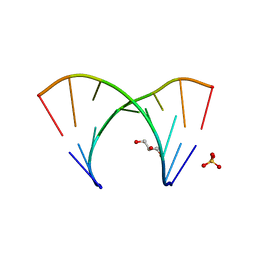
1.23 A resolution X-ray structure of (GCUGCUGC)2
分子名称: 5'-R(*GP*CP*UP*GP*CP*UP*GP*C)-3', GLYCEROL, SULFATE ION
著者Kiliszek, A, Kierzek, R, Krzyzosiak, W.J, Rypniewski, W.
登録日2009-03-12
公開日2009-05-26
最終更新日2024-04-03
実験手法X-RAY DIFFRACTION (1.23 Å)
主引用文献Structural insights into CUG repeats containing the 'stretched U-U wobble': implications for myotonic dystrophy.
Nucleic Acids Res., 37, 2009
2H6Y
DownloadVisualize
BU of 2h6y by Molmil
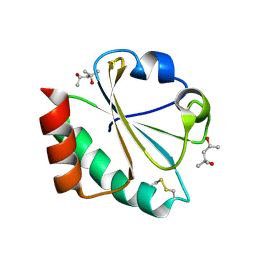
Crystal Structure of Thioredoxin Mutant E48D in Hexagonal (p61) Space Group
分子名称: (4S)-2-METHYL-2,4-PENTANEDIOL, Thioredoxin
著者Gavira, J.A, Godoy-Ruiz, R, Ibarra-Molero, B, Sanchez-Ruiz, J.M.
登録日2006-06-01
公開日2007-05-15
最終更新日2024-04-03
実験手法X-RAY DIFFRACTION (2.4 Å)
主引用文献Crystal Structure of Thioredoxin Mutant E48D in Hexagonal (p61) Space Group
To be Published
2FH9
DownloadVisualize
BU of 2fh9 by Molmil
Structure and dimerization of the kinase domain from yeast Snf1
分子名称: Snf1 kinase
著者Nayak, V.
登録日2005-12-23
公開日2006-03-28
最終更新日2024-02-14
実験手法X-RAY DIFFRACTION (2.8 Å)
主引用文献Structure and Dimerization of the Kinase Domain from Yeast Snf1, a Member of the Snf1/AMPK Protein Family
Structure, 14, 2006
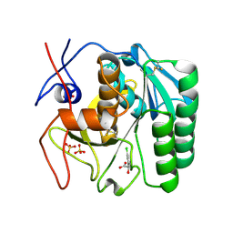
5ROP
DownloadVisualize
BU of 5rop by Molmil
PanDDA analysis group deposition -- Proteinase K changed state model for fragment Frag Xtal Screen A12a at Room Temperature
分子名称: (2R)-(3-chlorophenyl)(hydroxy)ethanoic acid, Proteinase K, SULFATE ION
著者Lima, G.M.A, Talibov, V, Benz, L.S, Jagudin, E, Mueller, U.
登録日2020-09-23
公開日2021-05-26
最終更新日2021-06-23
実験手法X-RAY DIFFRACTION (1.64 Å)
主引用文献FragMAXapp: crystallographic fragment-screening data-analysis and project-management system.
Acta Crystallogr D Struct Biol, 77, 2021
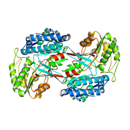
4I3U
DownloadVisualize
BU of 4i3u by Molmil
Structure of phosphonoacetaldehyde dehydrogenase in complex with phosphonoacetaldehyde
分子名称: Aldehyde dehydrogenase (NAD+), PHOSPHONOACETALDEHYDE
著者Nair, S.K, Agarwal, V.
登録日2012-11-26
公開日2013-11-27
最終更新日2019-07-17
実験手法X-RAY DIFFRACTION (2.1 Å)
主引用文献Structure and function of phosphonoacetaldehyde dehydrogenase: the missing link in phosphonoacetate formation.
Chem.Biol., 21, 2014
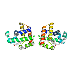
2QFK
DownloadVisualize
BU of 2qfk by Molmil
X-ray Crystal Structure Analysis of the Binding Site in the Ferric and Oxyferrous Forms of the Recombinant Heme Dehaloperoxidase Cloned from Amphitrite ornata
分子名称: AMMONIUM ION, Dehaloperoxidase A, PROTOPORPHYRIN IX CONTAINING FE, ...
著者de Serrano, V.S, Chen, Z, Davis, M.F, Franzen, S.
登録日2007-06-27
公開日2008-07-08
最終更新日2024-02-21
実験手法X-RAY DIFFRACTION (1.62 Å)
主引用文献X-ray crystal structural analysis of the binding site in the ferric and oxyferrous forms of the recombinant heme dehaloperoxidase cloned from Amphitrite ornata
Acta Crystallogr.,Sect.D, 63, 2007
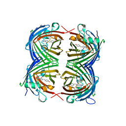
4HVF
DownloadVisualize
BU of 4hvf by Molmil
Crystal structure of green fluorescent protein lanGFP(Branchiostoma Lanceolatum)
分子名称: GLYCEROL, Green fluorescent protein blFP-Y6
著者Pletnev, V.Z, Pletneva, N.V, Pletnev, S.V.
登録日2012-11-06
公開日2013-09-18
最終更新日2023-12-06
実験手法X-RAY DIFFRACTION (1.7 Å)
主引用文献Structure of the red fluorescent protein from a lancelet (Branchiostoma lanceolatum): a novel GYG chromophore covalently bound to a nearby tyrosine.
Acta Crystallogr.,Sect.D, 69, 2013
3PVZ
DownloadVisualize
BU of 3pvz by Molmil
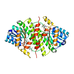
UDP-N-acetylglucosamine 4,6-dehydratase from Vibrio fischeri
分子名称: NICOTINAMIDE-ADENINE-DINUCLEOTIDE, SODIUM ION, THIOCYANATE ION, ...
著者Osipiuk, J, Marshall, N, Tesar, C, Pearson, L, Buck, K, Joachimiak, A, Midwest Center for Structural Genomics (MCSG)
登録日2010-12-07
公開日2011-01-12
最終更新日2017-11-08
実験手法X-RAY DIFFRACTION (2.1 Å)
主引用文献UDP-N-acetylglucosamine 4,6-dehydratase from Vibrio fischeri.
To be Published
3H6K
DownloadVisualize
BU of 3h6k by Molmil
Crystal Structure of Human 11-beta-hydroxysteroid-dehydrogenase Bound to an Ortho-chlro-sulfonyl-piperazine Inhibitor
分子名称: 3-chloro-4-({(2R)-4-[4-fluoro-2-(trifluoromethyl)phenyl]-2-methylpiperazin-1-yl}sulfonyl)benzamide, Corticosteroid 11-beta-dehydrogenase isozyme 1, NADP NICOTINAMIDE-ADENINE-DINUCLEOTIDE PHOSPHATE
著者Bard, J, Svenson, K.
登録日2009-04-23
公開日2009-12-01
最終更新日2024-02-21
実験手法X-RAY DIFFRACTION (2.187 Å)
主引用文献Efficacious 11beta-hydroxysteroid dehydrogenase type I inhibitors in the diet-induced obesity mouse model.
J.Med.Chem., 52, 2009
3PWT
DownloadVisualize
BU of 3pwt by Molmil
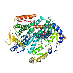
Crystal structure of mutant E.coli topoisomerase IA
分子名称: DNA topoisomerase, SULFATE ION
著者Zhang, Z, Bokun, C, Tse-Dinh, Y.-C.
登録日2010-12-08
公開日2010-12-29
最終更新日2024-02-21
実験手法X-RAY DIFFRACTION (1.9 Å)
主引用文献Crystal structure of mutant E.coli topoisomerase IA
To be Published
3PX0
DownloadVisualize
BU of 3px0 by Molmil
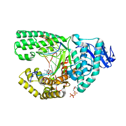
Crystal Structure of Bacillus DNA Polymerase I Large Fragment Bound to DNA and dCTP-dA Mismatch (tautomer) in Closed Conformation
分子名称: 2'-DEOXYCYTIDINE-5'-TRIPHOSPHATE, DNA (5'-D(*C*AP*TP*AP*GP*GP*AP*GP*TP*CP*AP*GP*G)-3'), DNA (5'-D(*CP*CP*TP*GP*AP*CP*TP*CP*(DOC))-3'), ...
著者Wang, W, Beese, L.S.
登録日2010-12-09
公開日2011-10-19
最終更新日2023-09-13
実験手法X-RAY DIFFRACTION (1.73 Å)
主引用文献Structural evidence for the rare tautomer hypothesis of spontaneous mutagenesis.
Proc.Natl.Acad.Sci.USA, 108, 2011
2QQM
DownloadVisualize
BU of 2qqm by Molmil
Crystal Structure of the a2b1b2 Domains from Human Neuropilin-1
分子名称: 1,2-ETHANEDIOL, 2-acetamido-2-deoxy-beta-D-glucopyranose-(1-4)-2-acetamido-2-deoxy-beta-D-glucopyranose, CALCIUM ION, ...
著者Appleton, B.A, Wiesmann, C.
登録日2007-07-26
公開日2007-11-20
最終更新日2023-08-30
実験手法X-RAY DIFFRACTION (2 Å)
主引用文献Structural studies of neuropilin/antibody complexes provide insights into semaphorin and VEGF binding
Embo J., 26, 2007
4HY2
DownloadVisualize
BU of 4hy2 by Molmil
Crystal structure of Plk1 Polo-box domain in complex with PL-42
分子名称: PL-42, Serine/threonine-protein kinase PLK1
著者Lee, W.C, Song, J.H, Kim, H.Y.
登録日2012-11-12
公開日2013-04-03
最終更新日2017-11-15
実験手法X-RAY DIFFRACTION (2 Å)
主引用文献Development of cyclic peptomer inhibitors targeting the polo-box domain of polo-like kinase 1.
Bioorg.Med.Chem., 21, 2013
2ESP
DownloadVisualize
BU of 2esp by Molmil
Human ubiquitin-conjugating enzyme (E2) UbcH5b mutant Ile88Ala
分子名称: (4S)-2-METHYL-2,4-PENTANEDIOL, CHLORIDE ION, Ubiquitin-conjugating enzyme E2 D2
著者Ozkan, E, Yu, H, Deisenhofer, J.
登録日2005-10-26
公開日2005-12-06
最終更新日2023-08-23
実験手法X-RAY DIFFRACTION (1.52 Å)
主引用文献Mechanistic insight into the allosteric activation of a ubiquitin-conjugating enzyme by RING-type ubiquitin ligases
Proc.Natl.Acad.Sci.Usa, 102, 2005

222624

件を2024-07-17に公開中

PDB statisticsPDBj update infoContact PDBjnumon