Loading
PDBj
メニューPDBj@FacebookPDBj@TwitterPDBj@YouTubewwPDB FoundationwwPDB
RCSB PDBPDBeBMRBAdv. SearchSearch help

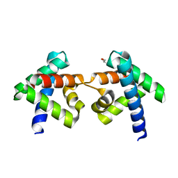
7DNS
DownloadVisualize
BU of 7dns by Molmil
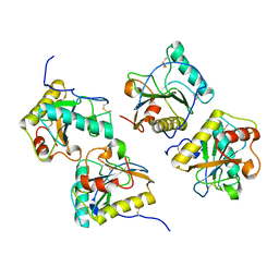
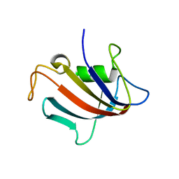
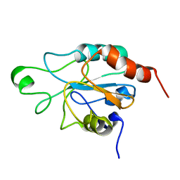
Crystal structure of domain-swapped dimer of H5_Fold-0 Elsa; de novo designed protein with an asymmetric all-alpha topology
分子名称: GLYCEROL, de novo designed protein
著者Suzuki, K, Kobayashi, N, Murata, T, Sakuma, K, Kosugi, T, Koga, R, Koga, N.
登録日2020-12-10
公開日2021-07-28
最終更新日2024-01-10
実験手法X-RAY DIFFRACTION (2.327 Å)
主引用文献Design of complicated all-alpha protein structures
Nat.Struct.Mol.Biol., 2024
7SH6
DownloadVisualize
BU of 7sh6 by Molmil
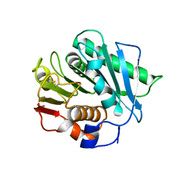
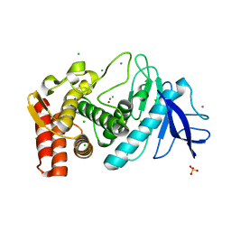
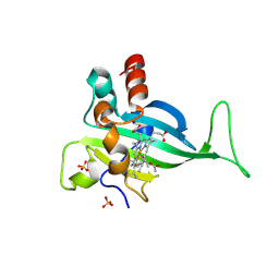
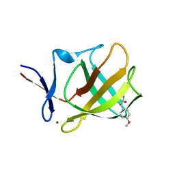
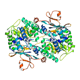
Crystal structure of a PET hydrolase mutant from Ideonella Sakaiensis
分子名称: Poly(ethylene terephthalate) hydrolase, SULFATE ION
著者Kim, W, Zhang, Y.
登録日2021-10-08
公開日2022-04-06
最終更新日2023-10-18
実験手法X-RAY DIFFRACTION (1.44 Å)
主引用文献Machine learning-aided engineering of hydrolases for PET depolymerization.
Nature, 604, 2022
2R5I
DownloadVisualize
BU of 2r5i by Molmil
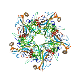
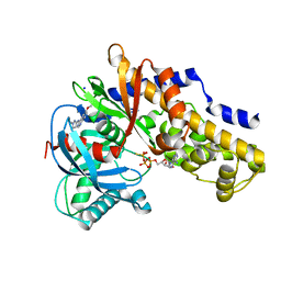
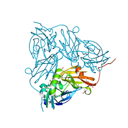
Pentamer Structure of Major Capsid Protein L1 of Human Papilloma Virus type 18
分子名称: L1 protein
著者Bishop, B, Dasgupta, J, Chen, X.S.
登録日2007-09-03
公開日2008-05-06
最終更新日2024-02-21
実験手法X-RAY DIFFRACTION (3.4 Å)
主引用文献Crystal structures of four types of human papillomavirus L1 capsid proteins: understanding the specificity of neutralizing monoclonal antibodies.
J.Biol.Chem., 282, 2007
2G2B
DownloadVisualize
BU of 2g2b by Molmil
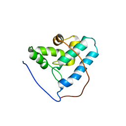
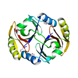
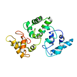
NMR structure of the human allograft inflammatory factor 1
分子名称: Allograft inflammatory factor 1
著者Song, J, Tyler, R.C, Newman, C.L, Vinarov, D, Markley, J.L, Center for Eukaryotic Structural Genomics (CESG)
登録日2006-02-15
公開日2006-02-28
最終更新日2024-05-29
実験手法SOLUTION NMR
主引用文献NMR structure of the human allograft inflammatory factor 1
To be published
2RTI
DownloadVisualize
BU of 2rti by Molmil
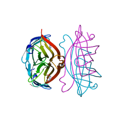
STREPTAVIDIN-GLYCOLURIL COMPLEX, PH 2.50, SPACE GROUP I222
分子名称: FORMIC ACID, GLYCOLURIL, STREPTAVIDIN
著者Katz, B.A.
登録日1997-09-11
公開日1998-10-14
最終更新日2024-02-21
実験手法X-RAY DIFFRACTION (1.4 Å)
主引用文献Binding of biotin to streptavidin stabilizes intersubunit salt bridges between Asp61 and His87 at low pH.
J.Mol.Biol., 274, 1997
2R90
DownloadVisualize
BU of 2r90 by Molmil
Crystal structure of cameline peptidoglycan recognition protein at 2.8A resolution
分子名称: Peptidoglycan recognition protein
著者Sharma, P, Singh, N, Sinha, M, Sharma, S, Kaur, P, Srinivasan, A, Singh, T.P.
登録日2007-09-12
公開日2007-09-25
最終更新日2023-10-25
実験手法X-RAY DIFFRACTION (2.8 Å)
主引用文献Crystal structure of cameline peptidoglycan recognition protein at 2.8A resolution
To be Published
2G4Z
DownloadVisualize
BU of 2g4z by Molmil
anomalous substructure of thermolysin
分子名称: CALCIUM ION, CHLORIDE ION, SULFATE ION, ...
著者Mueller-Dieckmann, C, Weiss, M.S.
登録日2006-02-22
公開日2007-02-20
最終更新日2024-02-14
実験手法X-RAY DIFFRACTION (1.98 Å)
主引用文献On the routine use of soft X-rays in macromolecular crystallography. Part IV. Efficient determination of anomalous substructures in biomacromolecules using longer X-ray wavelengths.
Acta Crystallogr.,Sect.D, 63, 2007
2RJI
DownloadVisualize
BU of 2rji by Molmil
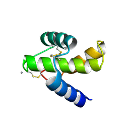
Malarial EBA-175 region VI crystallographic structure reveals a KIX-like binding interface
分子名称: CALCIUM ION, Erythrocyte binding antigen 175
著者Withers-Martinez, C, Blackman, M.J.
登録日2007-10-15
公開日2007-11-27
最終更新日2021-11-10
実験手法X-RAY DIFFRACTION (1.8 Å)
主引用文献Malarial EBA-175 Region VI Crystallographic Structure Reveals a KIX-Like Binding Interface
J.Mol.Biol., 375, 2008
2G69
DownloadVisualize
BU of 2g69 by Molmil
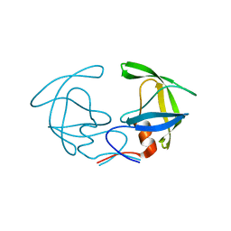
Structure of Unliganded HIV-1 Protease F53L Mutant
分子名称: protease
著者Kovalevsky, A.Y, Liu, F.
登録日2006-02-24
公開日2006-05-09
最終更新日2024-02-14
実験手法X-RAY DIFFRACTION (1.35 Å)
主引用文献Mechanism of Drug Resistance Revealed by the Crystal Structure of the Unliganded HIV-1 Protease with F53L Mutation.
J.Mol.Biol., 358, 2006
5I98
DownloadVisualize
BU of 5i98 by Molmil
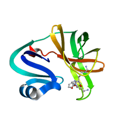
Structure of apo FKBP12(P104G) from C. albicans
分子名称: FK506-binding protein 1
著者Schumacher, M.A.
登録日2016-02-19
公開日2016-09-14
最終更新日2024-03-06
実験手法X-RAY DIFFRACTION (1.999 Å)
主引用文献Structures of Pathogenic Fungal FKBP12s Reveal Possible Self-Catalysis Function.
Mbio, 7, 2016
7S5O
DownloadVisualize
BU of 7s5o by Molmil
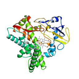
Crystal structure of Cytochrome c' beta from Nitrosomonas europaea ATCC 19718
分子名称: ACETATE ION, Cytochrome_P460 domain-containing protein, HEME C, ...
著者Seattle Structural Genomics Center for Infectious Disease (SSGCID)
登録日2021-09-11
公開日2022-03-30
最終更新日2022-04-20
実験手法X-RAY DIFFRACTION (1.8 Å)
主引用文献Structural Characterization of Cytochrome c ' beta-Met from an Ammonia-Oxidizing Bacterium.
Biochemistry, 61, 2022
3VEY
DownloadVisualize
BU of 3vey by Molmil
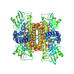
glucokinase in complex with glucose and ATPgS
分子名称: 6-methoxy-N-(1-methyl-1H-pyrazol-3-yl)quinazolin-4-amine, Glucokinase, PHOSPHOTHIOPHOSPHORIC ACID-ADENYLATE ESTER, ...
著者Liu, S.
登録日2012-01-09
公開日2012-02-08
最終更新日2024-02-28
実験手法X-RAY DIFFRACTION (2.25 Å)
主引用文献Insights into Mechanism of Glucokinase Activation: OBSERVATION OF MULTIPLE DISTINCT PROTEIN CONFORMATIONS.
J.Biol.Chem., 287, 2012
2RBB
DownloadVisualize
BU of 2rbb by Molmil
Crystal structure of a glyoxalase/bleomycin resistance protein/dioxygenase family enzyme from Burkholderia phytofirmans PsJN
分子名称: Glyoxalase/bleomycin resistance protein/dioxygenase
著者Rao, K.N, Sauder, J.M, Burley, S.K, Swaminathan, S, New York SGX Research Center for Structural Genomics (NYSGXRC)
登録日2007-09-18
公開日2007-10-16
最終更新日2021-10-20
実験手法X-RAY DIFFRACTION (1.82 Å)
主引用文献Crystal structure of a glyoxalase/bleomycin resistance protein/dioxygenase family enzyme from Burkholderia phytofirmans PsJN.
To be Published
2RM6
DownloadVisualize
BU of 2rm6 by Molmil
Glutathione peroxidase-type tryparedoxin peroxidase, reduced form
分子名称: Glutathione peroxidase-like protein
著者Melchers, J, Feher, K, Diechtierow, M, Krauth-Siegel, L, Muhle-Goll, C.
登録日2007-10-09
公開日2008-07-29
最終更新日2024-05-29
実験手法SOLUTION NMR
主引用文献Structural basis for a distinct catalytic mechanism in Trypanosoma brucei tryparedoxin peroxidase
J.Biol.Chem., 283, 2008
7SHH
DownloadVisualize
BU of 7shh by Molmil
Bacterial cereblon homologue in complex with (R)-3-(4-methoxyphenyl)piperidine-2,6-dione
分子名称: (3R)-3-(4-methoxyphenyl)piperidine-2,6-dione, Cereblon isoform 4, ZINC ION
著者Nithianantham, S, Fischer, M.
登録日2021-10-08
公開日2022-04-13
最終更新日2024-05-22
実験手法X-RAY DIFFRACTION (1.9 Å)
主引用文献Development of Potent and Selective Janus Kinase 2/3 Directing PG-PROTACs.
Acs Med.Chem.Lett., 13, 2022
5I6M
DownloadVisualize
BU of 5i6m by Molmil
Crystal Structure of Copper Nitrite Reductase at 100K after 7.59 MGy
分子名称: ACETATE ION, COPPER (II) ION, Copper-containing nitrite reductase, ...
著者Horrell, S, Hough, M.A, Strange, R.W.
登録日2016-02-16
公開日2016-07-13
最終更新日2024-01-10
実験手法X-RAY DIFFRACTION (1.09 Å)
主引用文献Serial crystallography captures enzyme catalysis in copper nitrite reductase at atomic resolution from one crystal.
Iucrj, 3, 2016
2G9B
DownloadVisualize
BU of 2g9b by Molmil
NMR solution structure of CA2+-loaded calbindin D28K
分子名称: Calbindin
著者Kojetin, D.J, Venters, R.A, Kordys, D.R, Thompson, R.J, Kumar, R, Cavanagh, J.
登録日2006-03-06
公開日2006-07-04
最終更新日2024-05-29
実験手法SOLUTION NMR
主引用文献Structure, binding interface and hydrophobic transitions of Ca(2+)-loaded calbindin-D(28K).
Nat.Struct.Mol.Biol., 13, 2006
2RN0
DownloadVisualize
BU of 2rn0 by Molmil
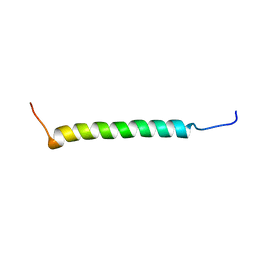
Micelle-embedded integrin beta3 transmembrane segment
分子名称: Integrin beta-3
著者Lau, T.L, Partridge, A.W, Ginsberg, M.H, Ulmer, T.S.
登録日2007-12-04
公開日2008-03-18
最終更新日2024-05-29
実験手法SOLUTION NMR
主引用文献Structure of the Integrin beta3 Transmembrane Segment in Phospholipid Bicelles and Detergent Micelles
Biochemistry, 47, 2008
2RNS
DownloadVisualize
BU of 2rns by Molmil
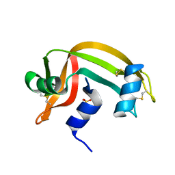
REFINEMENT OF THE CRYSTAL STRUCTURE OF RIBONUCLEASE S. COMPARISON WITH AND BETWEEN THE VARIOUS RIBONUCLEASE A STRUCTURES
分子名称: RIBONUCLEASE S, SULFATE ION
著者Kim, E.E, Varadarajan, R, Wyckoff, H.W, Richards, F.M.
登録日1992-02-19
公開日1994-01-31
最終更新日2019-08-14
実験手法X-RAY DIFFRACTION (1.6 Å)
主引用文献Refinement of the crystal structure of ribonuclease S. Comparison with and between the various ribonuclease A structures.
Biochemistry, 31, 1992
7SD1
DownloadVisualize
BU of 7sd1 by Molmil
Crystal structure of SHOC2
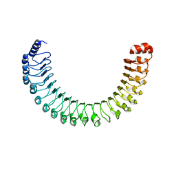
分子名称: Leucine-rich repeat protein SHOC-2
著者Liau, N.P.D, Hymowitz, S.G, Sudhamsu, J.
登録日2021-09-29
公開日2022-04-20
最終更新日2023-10-18
実験手法X-RAY DIFFRACTION (3.19 Å)
主引用文献Structural basis for SHOC2 modulation of RAS signalling.
Nature, 609, 2022
2RNW
DownloadVisualize
BU of 2rnw by Molmil
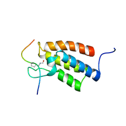
The Structural Basis for Site-Specific Lysine-Acetylated Histone Recognition by the Bromodomains of the Human Transcriptional Co-Activators PCAf and CBP
分子名称: Histone H3, Histone acetyltransferase PCAF
著者Zeng, L, Zhang, Q, Gerona-Navarro, G, Zhou, M.M.
登録日2008-02-03
公開日2008-05-06
最終更新日2023-11-15
実験手法SOLUTION NMR
主引用文献Structural Basis of Site-Specific Histone Recognition by the Bromodomains of Human Coactivators PCAF and CBP/p300
Structure, 16, 2008
2ROM
DownloadVisualize
BU of 2rom by Molmil
CRYSTAL STRUCTURE OF NITRIC REDUCTASE FROM DENITRIFYING FUNGUS FUSARIUM OXYSPORUM COMPLEX WITH CARBON MONOXIDE
分子名称: CARBON MONOXIDE, CYTOCHROME P450, PROTOPORPHYRIN IX CONTAINING FE
著者Park, S.-Y, Nakagawa, A.
登録日1997-03-24
公開日1997-10-15
最終更新日2024-02-21
実験手法X-RAY DIFFRACTION (2 Å)
主引用文献Crystal structure of nitric oxide reductase from denitrifying fungus Fusarium oxysporum.
Nat.Struct.Biol., 4, 1997
2G96
DownloadVisualize
BU of 2g96 by Molmil
Crystal Structure of Visfatin/Pre-B Cell Colony Enhancing Factor 1/Nicotinamide Phosphoribosyltransferase In Complex with Niconamide Mononucleotide
分子名称: BETA-NICOTINAMIDE RIBOSE MONOPHOSPHATE, Nicotinamide phosphoribosyltransferase
著者Eom, S.H, Kim, M.-K.
登録日2006-03-05
公開日2006-08-01
最終更新日2023-10-25
実験手法X-RAY DIFFRACTION (2.9 Å)
主引用文献Crystal Structure of Visfatin/Pre-B Cell Colony-enhancing Factor 1/Nicotinamide Phosphoribosyltransferase, Free and in Complex with the Anti-cancer Agent FK-866
J.Mol.Biol., 362, 2006
2REH
DownloadVisualize
BU of 2reh by Molmil
Mechanistic and Structural Analyses of the Roles of Arg409 and Asp402 in the Reaction of the Flavoprotein Nitroalkane Oxidase
分子名称: FLAVIN-ADENINE DINUCLEOTIDE, Nitroalkane oxidase
著者Fitzpatrick, P.F, Bozinovski, D.M, Heroux, A, Shaw, P.G, Valley, M.P, Orville, A.M.
登録日2007-09-26
公開日2008-06-03
最終更新日2023-10-25
実験手法X-RAY DIFFRACTION (2.4 Å)
主引用文献Mechanistic and structural analyses of the roles of Arg409 and Asp402 in the reaction of the flavoprotein nitroalkane oxidase.
Biochemistry, 46, 2007
7DA6
DownloadVisualize
BU of 7da6 by Molmil
Enterovirus 71 2A Protease mutant- C110A in complex with peptide inhibitor
分子名称: PHE-ARG-GLY-LYS, Polyprotein, ZINC ION
著者Yang, W.Z, Yuan, H.S.
登録日2020-10-15
公開日2021-08-25
最終更新日2023-11-29
実験手法X-RAY DIFFRACTION (2.2 Å)
主引用文献Efficient Strategy to Design Protease Inhibitors: Application to Enterovirus 71 2A Protease.
Acs Bio Med Chem Au, 2022

222624

件を2024-07-17に公開中

PDB statisticsPDBj update infoContact PDBjnumon