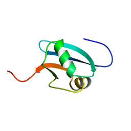
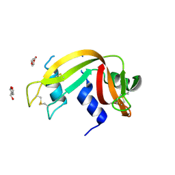
2UXN
 
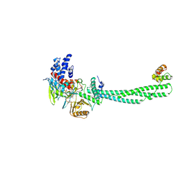 | | Structural Basis of Histone Demethylation by LSD1 Revealed by Suicide Inactivation | | 分子名称: | CHLORIDE ION, DIHYDROFLAVINE-ADENINE DINUCLEOTIDE, GLYCEROL, ... | | 著者 | Yang, M, Culhane, J.C, Szewczuk, L.M, Gocke, C.B, Brautigam, C.A, Tomchick, D.R, Machius, M, Cole, P.A, Yu, H. | | 登録日 | 2007-03-28 | | 公開日 | 2007-05-29 | | 最終更新日 | 2011-07-13 | | 実験手法 | X-RAY DIFFRACTION (2.72 Å) | | 主引用文献 | Structural Basis of Histone Demethylation by Lsd1 Revealed by Suicide Inactivation.
Nat.Struct.Mol.Biol., 14, 2007
|
|
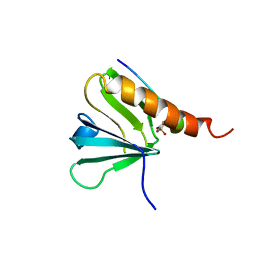
3EV3
 | | Crystal Structure of Ribonuclease A in 70% t-Butanol | | 分子名称: | Ribonuclease pancreatic, TERTIARY-BUTYL ALCOHOL | | 著者 | Dechene, M, Wink, G, Smith, M, Swartz, P, Mattos, C. | | 登録日 | 2008-10-12 | | 公開日 | 2009-06-23 | | 最終更新日 | 2023-12-27 | | 実験手法 | X-RAY DIFFRACTION (1.68 Å) | | 主引用文献 | Multiple solvent crystal structures of ribonuclease A: An assessment of the method
Proteins, 76, 2009
|
|
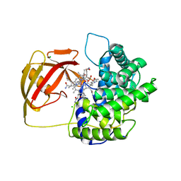
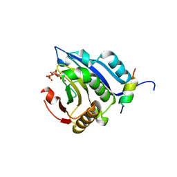
3EVR
 | | Crystal structure of Calcium bound monomeric GCAMP2 | | 分子名称: | CALCIUM ION, Myosin light chain kinase, Green fluorescent protein, ... | | 著者 | Wang, Q, Shui, B, Kotlikoff, M.I, Sondermann, H. | | 登録日 | 2008-10-13 | | 公開日 | 2008-12-09 | | 最終更新日 | 2023-12-27 | | 実験手法 | X-RAY DIFFRACTION (2 Å) | | 主引用文献 | Structural Basis for Calcium Sensing by GCaMP2.
Structure, 16, 2008
|
|
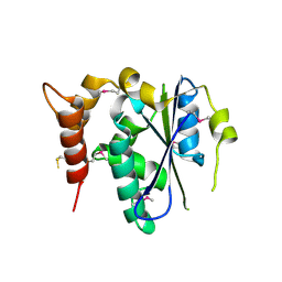
2UZ8
 | | The crystal structure of p18, human translation elongation factor 1 epsilon 1 | | 分子名称: | EUKARYOTIC TRANSLATION ELONGATION FACTOR 1 EPSILON-1, GLYCEROL | | 著者 | Kang, B.S, Kim, K.J, Kim, M.H, Oh, Y.S, Kim, S. | | 登録日 | 2007-04-26 | | 公開日 | 2008-03-25 | | 最終更新日 | 2011-07-13 | | 実験手法 | X-RAY DIFFRACTION (2 Å) | | 主引用文献 | Determination of Three-Dimensional Structure and Residues of the Novel Tumor Suppressor Aimp3/P18 Required for the Interaction with Atm.
J.Biol.Chem., 283, 2008
|
|
3ET1
 | |
3EGY
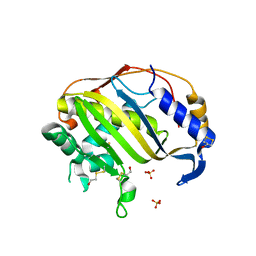 | | Crystal Structure of Human Thymidyalte Synthase A191K with Loop 181-197 stabilized in the inactive conformation | | 分子名称: | 1,2-ETHANEDIOL, SULFATE ION, Thymidylate synthase | | 著者 | Lovelace, L.L, Gibson, L.M, Johnson, S.R, Berger, S.H, Lebioda, L. | | 登録日 | 2008-09-11 | | 公開日 | 2009-08-18 | | 最終更新日 | 2023-08-30 | | 実験手法 | X-RAY DIFFRACTION (2.18 Å) | | 主引用文献 | Variants of human thymidylate synthase with loop 181-197 stabilized in the inactive conformation.
Protein Sci., 18, 2009
|
|
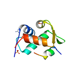
3EKJ
 | | Calcium-free GCaMP2 (calcium binding deficient mutant) | | 分子名称: | Myosin light chain kinase, Green fluorescent protein, Calmodulin chimera | | 著者 | Akerboom, J, Velez Rivera, J.D, Looger, L.L, Schreiter, E.R. | | 登録日 | 2008-09-19 | | 公開日 | 2008-12-16 | | 最終更新日 | 2023-11-15 | | 実験手法 | X-RAY DIFFRACTION (2.8 Å) | | 主引用文献 | Crystal Structures of the GCaMP Calcium Sensor Reveal the Mechanism of Fluorescence Signal Change and Aid Rational Design
J.Biol.Chem., 284, 2009
|
|
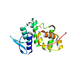
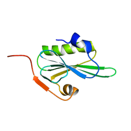
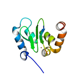
1J0G
 | | Solution Structure of Mouse Hypothetical 9.1 kDa Protein, A Ubiquitin-like Fold | | 分子名称: | Hypothetical Protein 1810045K17 | | 著者 | Zhao, C, Kigawa, T, Koshiba, S, Tochio, N, Kobayashi, N, Inoue, M, Yokoyama, S, RIKEN Structural Genomics/Proteomics Initiative (RSGI) | | 登録日 | 2002-11-13 | | 公開日 | 2003-12-09 | | 最終更新日 | 2023-12-27 | | 実験手法 | SOLUTION NMR | | 主引用文献 | Solution Structure of Mouse Hypothetical 9.1 kDa Protein, A Ubiquitin-like Fold
To be Published
|
|
2V3N
 | | Crystallographic analysis of upper axial ligand substitutions in cobalamin bound to transcobalamin | | 分子名称: | CHLORIDE ION, COBALAMIN, CYANIDE ION, ... | | 著者 | Wuerges, J, Geremia, S, Randaccio, L. | | 登録日 | 2007-06-19 | | 公開日 | 2007-10-30 | | 最終更新日 | 2023-12-13 | | 実験手法 | X-RAY DIFFRACTION (2.73 Å) | | 主引用文献 | Vitamin B12 Transport Proteins: Crystallographic Analysis of Beta-Axial Ligand Substitutions in Cobalamin Bound to Transcobalamin.
Iubmb Life, 59, 2007
|
|
3EQY
 | |
3EUY
 | | Crystal Structure of Ribonuclease A in 50% Dioxane | | 分子名称: | 1,4-DIETHYLENE DIOXIDE, Ribonuclease pancreatic | | 著者 | Dechene, M, Wink, G, Smith, M, Swartz, P, Mattos, C. | | 登録日 | 2008-10-12 | | 公開日 | 2009-06-23 | | 最終更新日 | 2023-12-27 | | 実験手法 | X-RAY DIFFRACTION (1.95 Å) | | 主引用文献 | Multiple solvent crystal structures of ribonuclease A: An assessment of the method
Proteins, 76, 2009
|
|
3EV6
 | | Crystal Structure of Ribonuclease A in 50% R,S,R-Bisfuranol | | 分子名称: | (3R,3aS,6aR)-hexahydrofuro[2,3-b]furan-3-ol, Ribonuclease pancreatic | | 著者 | Dechene, M, Wink, G, Smith, M, Swartz, P, Mattos, C. | | 登録日 | 2008-10-12 | | 公開日 | 2009-06-23 | | 最終更新日 | 2023-12-27 | | 実験手法 | X-RAY DIFFRACTION (1.76 Å) | | 主引用文献 | Multiple solvent crystal structures of ribonuclease A: An assessment of the method
Proteins, 76, 2009
|
|
1IRS
 | | IRS-1 PTB DOMAIN COMPLEXED WITH A IL-4 RECEPTOR PHOSPHOPEPTIDE, NMR, MINIMIZED AVERAGE STRUCTURE | | 分子名称: | IL-4 RECEPTOR PHOSPHOPEPTIDE, IRS-1 | | 著者 | Zhou, M.-M, Huang, B, Olejniczak, E.T, Meadows, R.P, Shuker, S.B, Miyazaki, M, Trub, T, Shoelson, S.E, Feisk, S.W. | | 登録日 | 1996-03-22 | | 公開日 | 1997-05-15 | | 最終更新日 | 2022-02-23 | | 実験手法 | SOLUTION NMR | | 主引用文献 | Structural basis for IL-4 receptor phosphopeptide recognition by the IRS-1 PTB domain.
Nat.Struct.Biol., 3, 1996
|
|
2V8W
 | | Crystallographic and mass spectrometric characterisation of eIF4E with N7-cap derivatives | | 分子名称: | EUKARYOTIC TRANSLATION INITIATION FACTOR 4E, EUKARYOTIC TRANSLATION INITIATION FACTOR 4E-BINDING PROTEIN 1, [[(2R,3S,4R,5R)-5-(6-AMINO-3-METHYL-4-OXO-5H-IMIDAZO[4,5-C]PYRIDIN-1-YL)-3,4-DIHYDROXY-OXOLAN-2-YL]METHOXY-HYDROXY-PHOSPHORYL] PHOSPHONO HYDROGEN PHOSPHATE | | 著者 | Brown, C.J, Mcnae, I, Fischer, P.M, Walkinshaw, M.D. | | 登録日 | 2007-08-16 | | 公開日 | 2007-08-28 | | 最終更新日 | 2023-12-13 | | 実験手法 | X-RAY DIFFRACTION (2.3 Å) | | 主引用文献 | Crystallographic and Mass Spectrometric Characterisation of Eif4E with N(7)-Alkylated CAP Derivatives.
J.Mol.Biol., 372, 2007
|
|
3ENP
 | | Crystal structure of human cgi121 | | 分子名称: | TP53RK-binding protein | | 著者 | Haffani, Y.Z, Ceccarelli, D.F, Neculai, D, Mao, D.Y, Sicheri, F. | | 登録日 | 2008-09-25 | | 公開日 | 2008-11-04 | | 最終更新日 | 2011-07-13 | | 実験手法 | X-RAY DIFFRACTION (2.48 Å) | | 主引用文献 | Atomic structure of the KEOPS complex: an ancient protein kinase-containing molecular machine.
Mol.Cell, 32, 2008
|
|
2VIK
 | |
3EUX
 | | Crystal Structure of Crosslinked Ribonuclease A | | 分子名称: | Ribonuclease pancreatic | | 著者 | Dechene, M, Wink, G, Smith, M, Swartz, P, Mattos, C. | | 登録日 | 2008-10-12 | | 公開日 | 2009-06-23 | | 最終更新日 | 2023-12-27 | | 実験手法 | X-RAY DIFFRACTION (1.65 Å) | | 主引用文献 | Multiple solvent crystal structures of ribonuclease A: An assessment of the method
Proteins, 76, 2009
|
|
3EV4
 | | Crystal Structure of Ribonuclease A in 50% Trifluoroethanol | | 分子名称: | Ribonuclease pancreatic, TRIFLUOROETHANOL | | 著者 | Dechene, M, Wink, G, Smith, M, Swartz, P, Mattos, C. | | 登録日 | 2008-10-12 | | 公開日 | 2009-06-23 | | 最終更新日 | 2023-12-27 | | 実験手法 | X-RAY DIFFRACTION (1.93 Å) | | 主引用文献 | Multiple solvent crystal structures of ribonuclease A: An assessment of the method
Proteins, 76, 2009
|
|
2V3E
 | | acid-beta-glucosidase with N-nonyl-deoxynojirimycin | | 分子名称: | (2R,3R,4R,5S)-2-(HYDROXYMETHYL)-1-NONYLPIPERIDINE-3,4,5-TRIOL, 2-acetamido-2-deoxy-beta-D-glucopyranose, GLUCOSYLCERAMIDASE, ... | | 著者 | Brumshtein, B, Greenblatt, H.M, Butters, T.D, Shaaltiel, Y, Aviezer, D, Silman, I, Futerman, A.H, Sussman, J.L. | | 登録日 | 2007-06-17 | | 公開日 | 2007-08-21 | | 最終更新日 | 2023-12-13 | | 実験手法 | X-RAY DIFFRACTION (2 Å) | | 主引用文献 | Crystal Structures of Complexes of N-Butyl- and N-Nonyl-Deoxynojirimycin Bound to Acid Beta-Glucosidase: Insights Into the Mechanism of Chemical Chaperone Action in Gaucher Disease.
J.Biol.Chem., 282, 2007
|
|
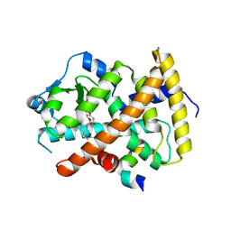
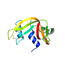
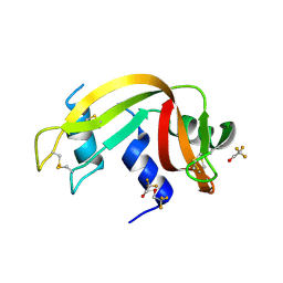
1JBH
 | | Solution structure of cellular retinol binding protein type-I in the ligand-free state | | 分子名称: | CELLULAR RETINOL-BINDING PROTEIN TYPE I | | 著者 | Franzoni, L, Luecke, C, Perez, C, Cavazzini, D, Rademacher, M, Ludwig, C, Spisni, A, Rossi, G.L, Rueterjans, H. | | 登録日 | 2001-06-04 | | 公開日 | 2002-06-19 | | 最終更新日 | 2024-05-22 | | 実験手法 | SOLUTION NMR | | 主引用文献 | Structure and backbone dynamics of Apo- and holo-cellular retinol-binding protein in solution.
J.Biol.Chem., 277, 2002
|
|
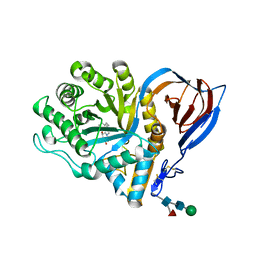
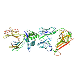
1JD6
 | | Crystal Structure of DIAP1-BIR2/Hid Complex | | 分子名称: | APOPTOSIS 1 INHIBITOR, ZINC ION, head involution defective protein | | 著者 | Wu, J.W, Cocina, A.E, Chai, J, Hay, B.A, Shi, Y. | | 登録日 | 2001-06-12 | | 公開日 | 2001-12-05 | | 最終更新日 | 2024-02-07 | | 実験手法 | X-RAY DIFFRACTION (2.7 Å) | | 主引用文献 | Structural analysis of a functional DIAP1 fragment bound to grim and hid peptides.
Mol.Cell, 8, 2001
|
|
2VLJ
 | | The Structural Dynamics and Energetics of an Immunodominant T-cell Receptor are Programmed by its Vbeta Domain | | 分子名称: | BETA-2-MICROGLOBULIN, FLU MATRIX PEPTIDE, HLA CLASS I HISTOCOMPATIBILITY ANTIGEN, ... | | 著者 | Ishizuka, J, Stewart-Jones, G, Van Der Merwe, A, Bell, J, Mcmichael, A, Jones, Y. | | 登録日 | 2008-01-15 | | 公開日 | 2008-01-22 | | 最終更新日 | 2011-07-13 | | 実験手法 | X-RAY DIFFRACTION (2.4 Å) | | 主引用文献 | The Structural Dynamics and Energetics of an Immunodominant T-Cell Receptor are Programmed by its Vbeta Domain
Immunity, 28, 2008
|
|
2VLL
 | | The Structural Dynamics and Energetics of an Immunodominant T-cell Receptor are Programmed by its Vbeta Domain | | 分子名称: | BETA-2-MICROGLOBULIN, FLU MATRIX PEPTIDE, HLA CLASS I HISTOCOMPATIBILITY ANTIGEN, ... | | 著者 | Ishizuka, J, Stewart-Jones, G, Van Der Merwe, A, Bell, J, Mcmichael, A, Jones, Y. | | 登録日 | 2008-01-15 | | 公開日 | 2008-01-22 | | 最終更新日 | 2011-07-13 | | 実験手法 | X-RAY DIFFRACTION (1.6 Å) | | 主引用文献 | The Structural Dynamics and Energetics of an Immunodominant T-Cell Receptor are Programmed by its Vbeta Domain
Immunity, 28, 2008
|
|
1JB6
 | |
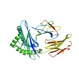
1JD4
 | | Crystal Structure of DIAP1-BIR2 | | 分子名称: | APOPTOSIS 1 INHIBITOR, ZINC ION | | 著者 | Wu, J.W, Cocina, A.E, Chai, J, Hay, B.A, Shi, Y. | | 登録日 | 2001-06-12 | | 公開日 | 2001-12-05 | | 最終更新日 | 2023-08-16 | | 実験手法 | X-RAY DIFFRACTION (2.7 Å) | | 主引用文献 | Structural analysis of a functional DIAP1 fragment bound to grim and hid peptides.
Mol.Cell, 8, 2001
|
|


